. 2000 May;74(9):4165-73.
doi: 10.1128/jvi.74.9.4165-4173.2000.

Intrahepatic induction of alpha/beta interferon eliminates viral RNA-containing capsids in hepatitis B virus transgenic mice

Affiliations

Intrahepatic induction of alpha/beta interferon eliminates viral RNA-containing capsids in hepatitis B virus transgenic mice

S F Wieland et al. J Virol. 2000 May.

Abstract

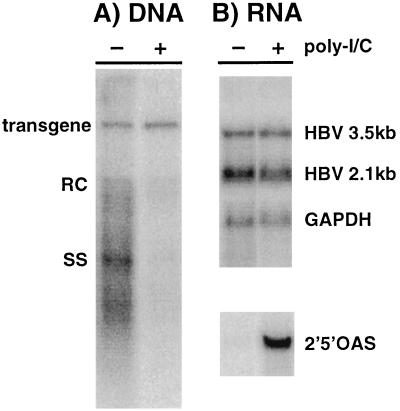
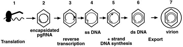
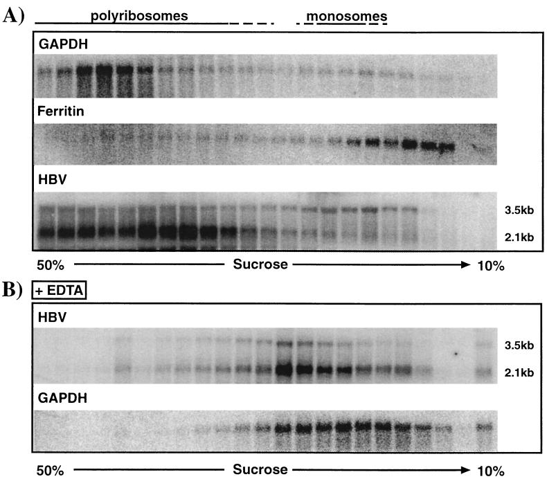
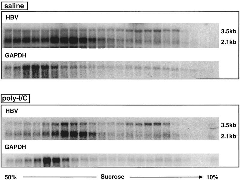
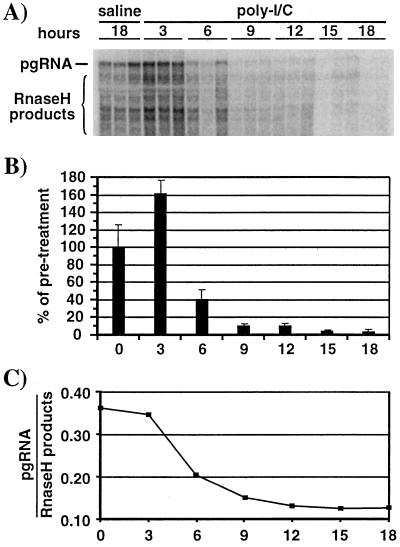
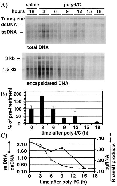
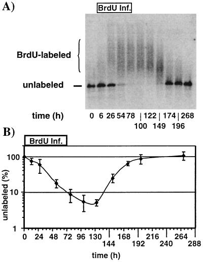
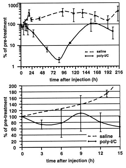
We have previously shown that hepatitis B virus (HBV) replication is abolished in the liver of HBV transgenic mice by stimuli that induce alpha/beta interferon (IFN-alpha/beta) in the liver. The present study was done to identify the step(s) in HBV replication that is affected by this cytokine in transgenic mice treated with the IFN-alpha/beta inducer polyinosinic-polycytidylic acid [poly(I-C)]. Here we show that the pool of cytoplasmic HBV pregenomic RNA (pgRNA)-containing capsids is reduced 10-fold within 9 h after poly(I-C) administration, while there is no change in the abundance of HBV mRNA or in the translational status of cytoplasmic HBV transcripts. In addition, we show that the pool of HBV DNA-containing capsids is not reduced to the same degree until at least 15 h posttreatment, and we show that virus export is not accelerated and the half-life of virions in the serum is unchanged. These results indicate that IFN-alpha/beta triggers intracellular events that either inhibit the assembly of pgRNA-containing capsids or accelerate their degradation, and that maturation and secretion of virus is responsible for clearance of HBV capsids and their cargo of replicative intermediates from the cytoplasm of the hepatocyte.


Figures

FIG. 1
FIG. 1
Poly(I-C) inhibits HBV replication but not gene expression in livers of transgenic mice. Two groups (three mice per group) of age (8 to 10 weeks)-, sex (male)-, and serum HBeAg-matched mice from lineage 1.3.32 were injected either with saline (−) or with a single i.v. dose (200 μg) of poly(I-C) (+). Twenty hours later, the mice were sacrificed; following extraction, total hepatic RNA and DNA were analyzed for HBV gene expression and replication by Northern and Southern blot analysis; a representative sample is shown. (A) Southern blot analysis was performed with 30 μg of total hepatic DNA. All DNA samples were RNase treated before gel electrophoresis. Bands corresponding to the integrated transgene, relaxed circular (RC), and single-stranded (SS) HBV DNA replicative forms are indicated. The integrated transgene can be used to normalize the amount of DNA bound to the membrane. The filter was hybridized with a 32P-labeled HBV-specific probe. (B) Northern blot analysis was performed with 20 μg of total hepatic RNA. The membrane was hybridized with 32P-labeled HBV-, 2′5′OAS-, and GAPDH-specific DNA probes. Bands corresponding to the 3.5- and 2.1-kb viral mRNAs are indicated.
FIG. 2
FIG. 2
Schematic representation of the posttranscriptional, cytoplasmic steps in the viral life cycle in livers of transgenic mice. Step 1, the HBV transcripts are translated into the viral gene products; step 2, viral pgRNA is encapsidated along with the viral RT/Pol; step 3, pgRNA is reverse transcribed by the RT/Pol and digested, leaving a short 5′ fragment and ssDNA of minus-strand polarity (step 4). Further capsid maturation involves transfer of the RNA fragment to the 3′ end of the ssDNA, where it serves as the primer for subsequent plus-strand DNA synthesis (step 5), which produces mature capsids containing dsDNA (step 6). Mature capsids are targeted for envelopment and are subsequently exported out of the cell as virions (step 7).
FIG. 3
FIG. 3
Polyribosome isolation from livers of HBV transgenic mice. Polyribosomes were harvested as described in Materials and Methods. (A) Serial fractions from sucrose gradients of polyribosomal liver extracts were analyzed by Northern blotting for the distribution of GAPDH, ferritin, and HBV 3.5- and 2.1-kb transcripts as described in Materials and Methods. (B) Polyribosomal liver extracts were treated with EDTA prior to sucrose density centrifugation, and fractions were analyzed by Northern blotting for HBV 3.5- and 2.1-kb transcripts and GAPDH transcripts as described above.
FIG. 4
FIG. 4
Poly(I-C) treatment does not alter the translational status of HBV transcripts. The polyribosomal distributions of HBV 3.5- and 2.1-kb and GAPDH transcripts in livers of HBV transgenic mice were analyzed 20 h after a single i.v. injection of saline or 200 μg of poly(I-C) as described in the legend to Fig. 3.
FIG. 5
FIG. 5
Poly(I-C) treatment eliminates pgRNA-containing capsids from the hepatocyte cytoplasm. Age (7 to 11 weeks)-, sex (male)-, and serum HBeAg-matched mice from lineage 1.3.32 were injected either with saline or with a single i.v. injection of poly(I-C). Groups of three mice were sacrificed at the indicated times after injection, and the liver tissue was harvested. (A) Northern blot analysis was performed to detect encapsidated viral RNA as described in Materials and Methods. RNA isolated from identical amounts of liver tissue (35 mg) was loaded. (B) Graphic representation of encapsidated pgRNA. Signals for the saline samples were set to 100%. (C) Ratio of encapsidated pgRNA versus RNase H products plotted for each time point after poly(I-C) injection. Signals were quantified from the indicated band (pgRNA) and region (RNase H products) shown in the Northern blot above. Saline samples were considered to be preinjection (time zero) samples. The mean values for each group of mice are shown.
FIG. 6
FIG. 6
ssDNA-containing capsids are cleared after RNA-containing capsids but before mature capsids. Liver tissue samples from the mice represented in Fig. 5 were processed as follows. (A) Southern blot analysis was performed with 30 μg of total liver DNA as described in the legend to Fig. 2 (top), and encapsidated DNA from the equivalent of 5 mg of liver tissue was extracted and analyzed by Southern blotting as described in the Materials and Methods (bottom). Bands corresponding to the integrated transgene, dsDNA, and ssDNA HBV DNA replicative forms and size markers are indicated. (B) Graphic representation of HBV DNA at the indicated time points after poly(I-C) injection. Bars represent the combined signals for dsDNA and ssDNA. Mean values for each group of mice are shown. The value for the saline sample was set to 100%. (C) Ratios of ssDNA versus dsDNA were plotted at each time point after poly(I-C) injection (solid line). Ratios of encapsidated pgRNA versus RNase H products from Fig. 5C were also included (dotted line). HBV DNA signals were quantified by phosphorimaging analysis of the Southern blot shown in the top part of panel A. Signals were corrected for variations in the transgene.
FIG. 7
FIG. 7
Determination of the viral half-life in sera of HBV transgenic mice. A group of four age (7 to 11 weeks)-, sex (male)-, and HBeAg-matched mice from lineage 1.3.46 were infused with BrdU as described in Materials and Methods. Serum samples were collected at the indicated time points. (A) Representative HBV-specific Southern blot analysis of a gel retardation experiment as described in Materials and Methods. Positions of unlabeled and BrdU-labeled DNA are indicated. (B) Levels of unlabeled serum HBV DNA were plotted on a log scale for each time point during BrdU infusion (0 to 72 h) and thereafter. The mean values of four mice are shown. Serum HBV DNA levels prior to BrdU infusion were set to 100%.
FIG. 8
FIG. 8
Poly(I-C) does not affect virus export or half-life. Two groups of age (6 to 8 weeks)-, sex (male)-, and serum HBeAg-matched mice from the 1.3.46 lineage were injected either with saline (two mice) or with a single i.v. injection of poly(I-C) (three mice), and serum samples were analyzed at different time points after injection. Serum HBV DNA levels were plotted on a log scale for all time points measured after poly(I-C) injection (top). The serum HBV DNA levels up to 15 h after poly(I-C) injection were replotted on a linear scale (bottom). Mean values for relative serum HBV DNA levels in each group were plotted. Pretreatment serum HBV DNA levels were set to 100%.

References

    1. Aziz N, Munro H N. Both subunits of rat liver ferritin are regulated at a translational level by iron induction. Nucleic Acids Res. 1986;14:915–927. - PMC - PubMed
    1. Carroll S S, Chen E, Viscount T, Geib J, Sardana M K, Gehman J, Kuo L C. Cleavage of oligoribonucleotides by the 2′,5′-oligoadenylate-dependent ribonuclease L. J Biol Chem. 1996;271:4988–4992. - PubMed
    1. Cavanaugh V J, Guidotti L G, Chisari F V. Inhibition of hepatitis B virus replication during adenovirus and cytomegalovirus infections in transgenic mice. J Virol. 1998;72:2630–2637. - PMC - PubMed
    1. Cavanaugh V J, Guidotti L G, Chisari F V. Interleukin-12 inhibits hepatitis B virus replication in transgenic mice. J Virol. 1997;71:3236–3243. - PMC - PubMed
    1. Chatterjee S, Hunter E, Whitley R. Effect of cloned human interferons on protein synthesis and morphogenesis of herpes simplex virus. J Virol. 1985;56:419–425. - PMC - PubMed

Publication types

MeSH terms

LinkOut - more resources