. 2000 May;74(9):4192-206.
doi: 10.1128/jvi.74.9.4192-4206.2000.

Dysregulation of cyclin E gene expression in human cytomegalovirus-infected cells requires viral early gene expression and is associated with changes in the Rb-related protein p130

Affiliations

Dysregulation of cyclin E gene expression in human cytomegalovirus-infected cells requires viral early gene expression and is associated with changes in the Rb-related protein p130

A K McElroy et al. J Virol. 2000 May.

Abstract

We have previously shown that many cell cycle regulatory gene products are markedly affected by infection of primary fibroblasts with human cytomegalovirus (HCMV) (F. M. Jault, J. M. Jault, F. Ruchti, E. A. Fortunato, C. Clark, J. Corbeil, D. D. Richman, and D. H. Spector, J. Virol. 69:6697-6704, 1995). One of these proteins, cyclin E, is a key determinant of cell cycle progression during G(1), and its mRNA levels are significantly increased in HCMV-infected fibroblasts (B. S. Salvant, E. A. Fortunato, and D. H. Spector, J. Virol. 72:3729-3741, 1998). To determine the molecular basis of this effect, we have examined the events that occur at the endogenous cyclin E promoter during the course of infection. In vivo dimethyl sulfate footprinting of the cyclin E promoter revealed several regions of protection and hypersensitivity that were unique to infected cells. In accord with this observation, we find that the virus-induced cyclin E transcripts initiate downstream of the start site identified in mock-infected cells, in regions where these newly appearing protected and hypersensitive sites occur. Viral gene expression is required for this induction. However, the viral immediate-early proteins IE1-72 and IE2-86, either alone or in combination, cannot induce expression of the endogenous cyclin E. The virus must progress past the immediate-early phase and express an early gene product(s) for activation of cyclin E expression. Moreover, IE1-72 does not appear to be required, as infection of cells with an HCMV mutant containing a deletion in the IE1-72 gene leads to full upregulation of cyclin E expression. Using electrophoretic mobility shift assays with infected cell extracts and a region of the cyclin E promoter that includes two previously defined E2F sites as the probe, we detected the appearance of an infection-specific banding pattern. One of the infection-specific bands contained the proteins E2F-4, DP-1, and p130, which were maintained in the infected cells as uniquely phosphorylated species. These results suggest that an altered E2F-4-DP-1-p130 complex along with viral early gene expression may play a role in the transcriptional regulation of cyclin E mRNA during HCMV infection.


Figures
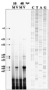
FIG. 1
FIG. 1
Primer extension analysis of cyclin E mRNA at 10 and 48 h p.i. mRNA was isolated using Invitrogen FastTrack 2.0, and primer extension was performed as previously described (49). Arrows indicate locations of transcription start sites relative to the published sequence (45). The site at +1 is marked to indicate the start site that was previously identified by Ohtani et al. (45). We do not detect use of this site in fibroblasts. Sanger sequencing reactions using the same primer as was used in the primer extension were performed in parallel. M, mock-infected cells; V, virus-infected cells.
FIG. 2
FIG. 2
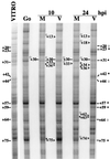
(Part 1) In vivo DMS footprinting analysis of the cyclin E promoter at 10 and 24 h p.i. Cells were treated with 0.1% DMS as described in Materials and Methods. After LMPCR, samples were run out on a 6% denaturing polyacrylamide gel. Hypersensitive sites are indicated by asterisks, and protected sites are identified by open circles. Lines denote changes that were seen in all in vivo samples compared to the in vitro sample. Arrows mark differences between mock (M)- and HCMV (V)-infected samples. In all cases, the corresponding location within the sequence is noted. The lane labeled G0 represents in vivo DMS treatment of confluent uninfected cells. The lane labeled VITRO represents in vitro treatment of DNA from uninfected cells. (Part 2) A summary of the events that occur on the cyclin E promoter. Solid arrows represent virus-induced transcription start sites. The open arrow designates the cellular start site for transcription. Solid asterisks denote hypersensitive sites that were detected in the in vivo footprint of infected cells. Solid circles mark protected sites from the in vivo footprint of infected cells. Open asterisks represent hypersensitivity that was seen in mock-infected cells, and open circles represent protection that was seen in mock-infected cells. The E2F site that is underlined in this panel is a component of the probe that was used in EMSAs.
FIG. 2
FIG. 2
(Part 1) In vivo DMS footprinting analysis of the cyclin E promoter at 10 and 24 h p.i. Cells were treated with 0.1% DMS as described in Materials and Methods. After LMPCR, samples were run out on a 6% denaturing polyacrylamide gel. Hypersensitive sites are indicated by asterisks, and protected sites are identified by open circles. Lines denote changes that were seen in all in vivo samples compared to the in vitro sample. Arrows mark differences between mock (M)- and HCMV (V)-infected samples. In all cases, the corresponding location within the sequence is noted. The lane labeled G0 represents in vivo DMS treatment of confluent uninfected cells. The lane labeled VITRO represents in vitro treatment of DNA from uninfected cells. (Part 2) A summary of the events that occur on the cyclin E promoter. Solid arrows represent virus-induced transcription start sites. The open arrow designates the cellular start site for transcription. Solid asterisks denote hypersensitive sites that were detected in the in vivo footprint of infected cells. Solid circles mark protected sites from the in vivo footprint of infected cells. Open asterisks represent hypersensitivity that was seen in mock-infected cells, and open circles represent protection that was seen in mock-infected cells. The E2F site that is underlined in this panel is a component of the probe that was used in EMSAs.
FIG. 3
FIG. 3
Western blot analysis of the E2F family of proteins at various times p.i. One hundred micrograms of total cell lysate was run on a 20-cm 10% polyacrylamide gel (0.07% bisacrylamide final concentration) (31). Proteins were transferred to nitrocellulose and analyzed by Western blotting. (A) E2F-1. (B) E2F-2. (C) E2F-3. (D) E2F-4. (E) E2F-5. (F) Nuclear and cytoplasmic lysates were prepared at 0 and 8 h p.i., and 100 μg of nuclear (N), cytoplasmic (C), or nuclear lysates treated with CIAP were run on 10% polyacrylamide gels. Proteins were transferred to nitrocellulose and analyzed by Western blotting for E2F-4. Two different exposures of the same blot are shown. (G) One hundred micrograms of nuclear (N) or cytoplasmic (C) lysates from three independent experiments was run on 10% polyacrylamide gels. Proteins were transferred to nitrocellulose and analyzed by Western blotting for G6PD and CBP. M, mock infected; V, virus infected.
FIG. 4
FIG. 4
Western blot analysis of other proteins that might be involved in transcriptional regulation of cyclin E. Lysates were prepared at various times p.i., and 100 μg of total cell lysate was used for each lane. Samples for DP-1 (A) and DP-2 (B) detection were run on a 20-cm 10% polyacrylamide gel (0.07% bisacrylamide final concentration) (31), transferred to nitrocellulose, and analyzed by Western blotting. (C) Samples for Sp-1 detection were run on a 10% minigel (29:1 bis/acrylamide ratio), transferred to Immobilon, and analyzed by Western blotting. Samples for p130 (D) and p107 (E) detection were run on a 7.5% polyacrylamide gel (0.07% bisacrylamide final concentration) (31), transferred to nitrocellulose, and analyzed by Western blotting.
FIG. 5
FIG. 5
EMSAs using sequences from the cyclin E promoter. (A) Nuclear extracts were prepared at various times p.i. Five micrograms of nuclear extract was combined with the radiolabeled probe CYCE E2F DS (5′ CT AGC GCC GGT TCC GCG CGC AGG GAT TTT AAA TGT CCC GCT CTG AG 3′) as described in Materials and Methods. The letter E designates the early virus- induced complex. The letter L designates the late virus-induced complex. The letter R designates the Rb-containing band shift. The letters NS designate nonspecific complexes. M, mock infected; V, virus infected. (B) Five micrograms of nuclear extract from infected cells at 18 h p.i. was combined with the same radiolabeled probe as in panel A. The indicated amount of competitor probe in molar excess was added to the reaction. The sequences of the competitor probes were as follows: FLAG, 5′ GAT CTA TGG ACT ACA AGG ACG ACG ACG ACA AGG G 3′; CYCE DS MUT, 5′ GCC GGT TCC GAT CGC AGG GAT TTT AAA TGT CAT GCT CTG AG 3′; DHFR E2F, 5′ GGG CGG GGC GGC CAC AAT TTC GCG CCA AAC TTG ACC GCG CG 3′; CYCA CDE/CHR, 5′ CCA TTT CAA TAG TCG CGG GAT ACT TGA ACT GCA AGA ACA GC 3′; and E2F CONS, 5′ GAT CTA TGG ATT TAA GTT TCG CGC CCT TTC TCA TAC TA 3′.
FIG. 6
FIG. 6
Comparison of the E2F complexes that are present in mock- and HCMV-infected cells. (A) The DHFR probe was radiolabeled and combined with 5 μg of nuclear lysate obtained from mock-infected cells at 24 h p.i. The indicated antiserum was added to the reaction to determine if the band shifts could be supershifted or abrogated by the specific antiserum. (B) The CYCE E2F DS probe was radiolabeled and combined with 5 μg of nuclear lysate obtained from HCMV-infected cells at 18 h p.i. The indicated antiserum was added to the reaction to determine if complex E could be supershifted or abrogated by the specific antiserum. The supershift that was produced by the addition of p130 antibody is marked by an arrow. A portion of the gel was cropped and the contrast was adjusted in order to better visualize the effects of the antiserum.
FIG. 7
FIG. 7
In vivo association between p130 and E2F-4. (A) Mock-infected cell lysates (M) from 12 h p.i. were immunoprecipitated (IP) with p130 antibody or rabbit preimmune serum coupled to protein A-Sepharose. IgG, immunoglobulin G. (B) Mock-infected (M) or infected (V) cell lysates from 8 and 24 h p.i. were immunoprecipitated (IP) with p130 antibody coupled to protein A-Sepharose. Immunoprecipitates were run on a 7.5% polyacrylamide gel. Proteins were transferred to nitrocellulose and Western blotted for p130 and E2F-4.
FIG. 8
FIG. 8
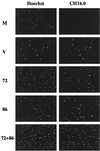
Recombinant baculoviruses express IE1-72 and IE2-86 in human fibroblasts. Cells were confluence synchronized and mock infected (M), infected with HCMV (V), or infected with recombinant baculovirus(es) (72, 86, or 72+86). Coverslips were taken at 24 h p.i., fixed and permeabilized in methanol, and stained with Hoechst stain to identify the total number of cells and CH16.0 to detect cells that expressed IE1-72 or IE2-86.
FIG. 9
FIG. 9
Expression of HCMV IE1-72 and that of IE2-86 are not sufficient to induce endogenous cyclin E. (A) Lysates were prepared at 24 h p.i. from the same experiment that was described in the legend to Fig. 8, and 50 μg of total cell lysate was run on a 10% polyacrylamide gel, transferred to nitrocellulose, and Western blotted for cyclin E. M, mock infected; V, virus infected. (B) Cells were serum starved and were mock infected (M), infected with HCMV (V), infected with UV-irradiated virus (UV), mock infected and infected with recombinant baculovirus (M and 72 or 86), or infected with both UV-irradiated virus and recombinant baculovirus (UV and 72 or 86). Cell lysates were prepared at 24 h p.i., and 50 μg of total cell lysate was run on a 10% polyacrylamide gel, transferred to nitrocellulose, and Western blotted for cyclin E.
FIG. 10
FIG. 10
IE1-72 is not required for the HCMV-mediated upregulation of cyclin E. Cells were serum starved and were mock infected (M), infected with RC303ΔAcc (Δ72), or infected with the Towne strain (V). Cell lysates were prepared at 24 h p.i., and 100 μg of total cell lysate was run on a 10% polyacrylamide gel, transferred to nitrocellulose, and Western blotted for HCMV IE1-72–IE2-86 and cyclin E.
FIG. 11
FIG. 11
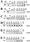
Viral early gene expression is required for the HCMV-mediated upregulation of cyclin E. Cells were confluence synchronized and mock (M)- or HCMV (V)-infected in the presence of cycloheximide (CHX). Cells were released from the cycloheximide block at 3 h p.i., and actinomycin D (ActD) was added at various times postrelease (0 to 15 h p.i.). Lysates were prepared at 18 h p.i., and 50 μg of total cell lysate was run on a 10% polyacrylamide gel, transferred to nitrocellulose, and Western blotted for IE1-72 and IE2-86 (CH16.0) (A and D), UL44 (B and E), or cyclin E (C and F). It should be noted that the apparent small increase in cyclin E in the mock-infected cells in panel F, lane 5, is due to the presence of BSA (due to inefficient washing of the cells prior to lysis) that comigrated at this position and nonspecifically bound some of the cyclin E antibody.

Similar articles

Cited by

References

    1. Ausubel F M, et al. Current protocols in molecular biology. New York, N.Y: Greene Publishing Associates and Wiley-Interscience; 1987.
    1. Beijersbergen R L, Bernards R. Cell cycle regulation by the retinoblastoma family of growth inhibitory proteins. Biochim Biophys Acta. 1996;1287:103–120. - PubMed
    1. Bresnahan W A, Albrecht T, Thompson E A. The cyclin E promoter is activated by human cytomegalovirus 86-kDa immediate early protein. J Biol Chem. 1998;273:22075–22082. - PubMed
    1. Bresnahan W A, Boldogh I, Chi P, Thompson E A, Albrecht T. Inhibition of cellular Cdk2 activity blocks human cytomegalovirus replication. Virology. 1997;231:239–247. - PubMed
    1. Bresnahan W A, Boldogh I, Thompson E A, Albrecht T. Human cytomegalovirus inhibits cellular DNA synthesis and arrests productively infected cells in late G1. Virology. 1996;224:150–160. - PubMed

Publication types

MeSH terms

Substances

LinkOut - more resources