

Apoptosis in coxsackievirus B3-caused diseases: interaction between the capsid protein VP2 and the proapoptotic protein siva
- PMID: 10756043
- PMCID: PMC111945
- DOI: 10.1128/jvi.74.9.4284-4290.2000
Apoptosis in coxsackievirus B3-caused diseases: interaction between the capsid protein VP2 and the proapoptotic protein siva
Abstract
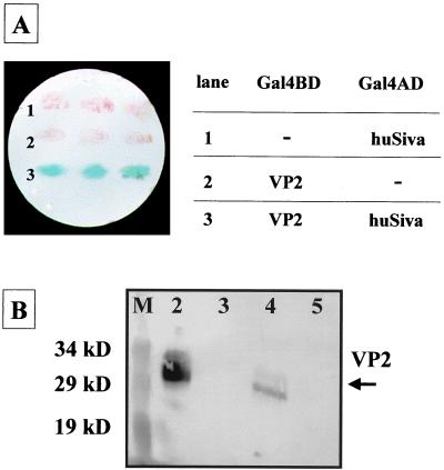
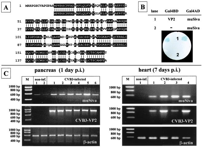
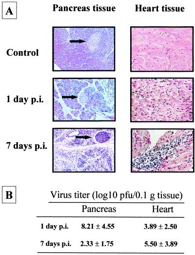
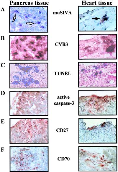
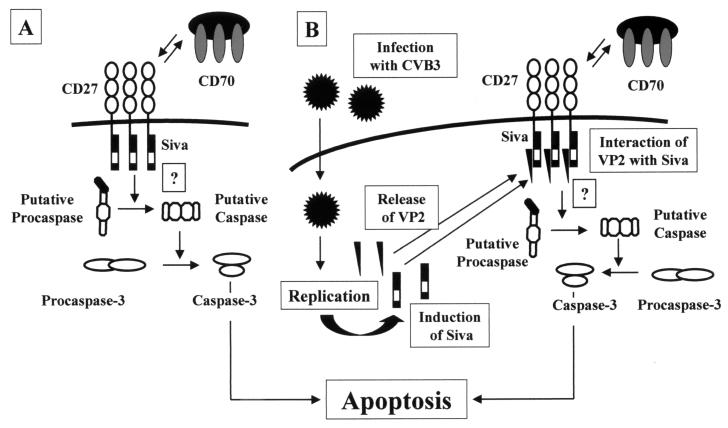
Coxsackievirus B3 (CVB3) is a common factor in human myocarditis. Apoptotic events are present in CVB3-induced disease, but it is unclear how CVB3 is involved in apoptosis and which viral proteins may induce the apoptotic pathway. In this report we demonstrate that the human and murine proapoptotic protein Siva specifically interact with the CVB3 capsid protein VP2. Furthermore, the transcription of Siva is strongly induced in tissue of CVB3-infected mice and is present in the same area which is positively stained for apoptosis, CD27, and CD70. It has been proposed that Siva is involved in the CD27/CD70-transduced apoptosis. Therefore, we suggest a molecular mechanism through which apoptotic events contributes to CVB3-caused pathogenesis.
Figures
Similar articles
-
The apoptotic capability of coxsackievirus B3 is influenced by the efficient interaction between the capsid protein VP2 and the proapoptotic host protein Siva.Virology. 2001 Oct 10;289(1):15-22. doi: 10.1006/viro.2001.1082. Virology. 2001. PMID: 11601913
-
A mutation in the puff region of VP2 attenuates the myocarditic phenotype of an infectious cDNA of the Woodruff variant of coxsackievirus B3.J Virol. 1996 Nov;70(11):7811-8. doi: 10.1128/JVI.70.11.7811-7818.1996. J Virol. 1996. PMID: 8892902 Free PMC article.
-
Proapoptotic protein Siva binds to the muscle protein telethonin in cardiomyocytes during coxsackieviral infection.Cardiovasc Res. 2009 Jan 1;81(1):108-15. doi: 10.1093/cvr/cvn276. Epub 2008 Oct 11. Cardiovasc Res. 2009. PMID: 18849585
-
A Kidnapping Story: How Coxsackievirus B3 and Its Host Cell Interact.Cell Physiol Biochem. 2019;53(1):121-140. doi: 10.33594/000000125. Cell Physiol Biochem. 2019. PMID: 31230428 Review.
-
Coxsackie B virus and its interaction with permissive host cells.Clin Diagn Virol. 1998 Apr;9(2-3):115-23. doi: 10.1016/s0928-0197(98)00010-5. Clin Diagn Virol. 1998. PMID: 9645993 Review.
Cited by
-
A single coxsackievirus B2 capsid residue controls cytolysis and apoptosis in rhabdomyosarcoma cells.J Virol. 2010 Jun;84(12):5868-79. doi: 10.1128/JVI.02383-09. Epub 2010 Apr 7. J Virol. 2010. PMID: 20375176 Free PMC article.
-
Gamma interferon-inducible protein 10 induces HeLa cell apoptosis through a p53-dependent pathway initiated by suppression of human papillomavirus type 18 E6 and E7 expression.Mol Cell Biol. 2005 Jul;25(14):6247-58. doi: 10.1128/MCB.25.14.6247-6258.2005. Mol Cell Biol. 2005. PMID: 15988033 Free PMC article.
-
Nicotinic Agonist Inhibits Cardiomyocyte Apoptosis in CVB3-Induced Myocarditis via α3β4-nAChR/PI3K/Akt-Dependent Survivin Upregulation.Oxid Med Cell Longev. 2019 Mar 7;2019:9496419. doi: 10.1155/2019/9496419. eCollection 2019. Oxid Med Cell Longev. 2019. PMID: 30984342 Free PMC article.
-
Interaction of CSFV E2 protein with swine host factors as detected by yeast two-hybrid system.PLoS One. 2014 Jan 8;9(1):e85324. doi: 10.1371/journal.pone.0085324. eCollection 2014. PLoS One. 2014. PMID: 24416391 Free PMC article.
-
Coxsackievirus B3 replication is reduced by inhibition of the extracellular signal-regulated kinase (ERK) signaling pathway.J Virol. 2002 Apr;76(7):3365-73. doi: 10.1128/jvi.76.7.3365-3373.2002. J Virol. 2002. PMID: 11884562 Free PMC article.
References
-
- Agol V I, Belov G A, Bienz K, Egger D, Kolesnikova M S, Raikhlin N T, Romanova L I, Smirnova E A, Tolskaya E A. Two types of death of poliovirus-infected cells: caspase involvement in the apoptosis but not cytopathic effect. Virology. 1998;252:343–353. - PubMed
-
- Badorff C, Lee G H, Lamphear B J, Martone M E, Campbell K P, Rhoads R E, Knowlton K U. Enteroviral protease 2A cleaves dystrophin: evidence of cytoskeletal disruption in an acquired cardiomyopathy. Nat Med. 1999;5:320–326. - PubMed
-
- Barco A, Carrasco L. Poliovirus 2Apro expression inhibits growth of yeast cells. FEBS Lett. 1995;371:4–8. - PubMed
-
- Bartel P, Chien C T, Sternglanz R, Fields S. Elimination of false positives that arise in using the two-hybrid system. BioTechniques. 1993;14:920–924. - PubMed
-
- Bergelson J M, Cunningham J A, Droguett G, Kurt-Jones E A, Krithivas A, Hong J S, Horwitz M S, Crowell R L, Finberg R W. Isolation of a common receptor for coxsackie B viruses and adenoviruses 2 and 5. Science. 1997;275:1320–1323. - PubMed
Publication types
MeSH terms
Substances
LinkOut - more resources
Full Text Sources
Other Literature Sources
Molecular Biology Databases
Research Materials
