. 2000 May;74(9):4291-301.
doi: 10.1128/jvi.74.9.4291-4301.2000.

Virus-specific cofactor requirement and chimeric hepatitis C virus/GB virus B nonstructural protein 3

Affiliations

Virus-specific cofactor requirement and chimeric hepatitis C virus/GB virus B nonstructural protein 3

N Butkiewicz et al. J Virol. 2000 May.

Abstract

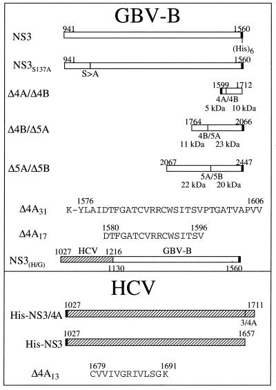
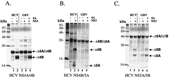
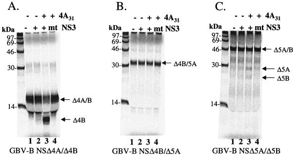
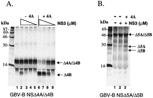
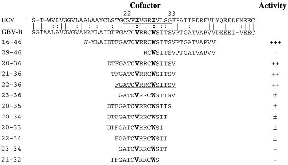
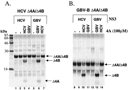
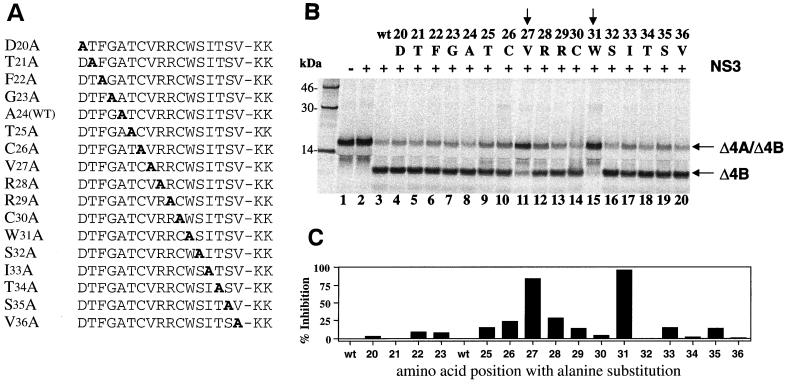
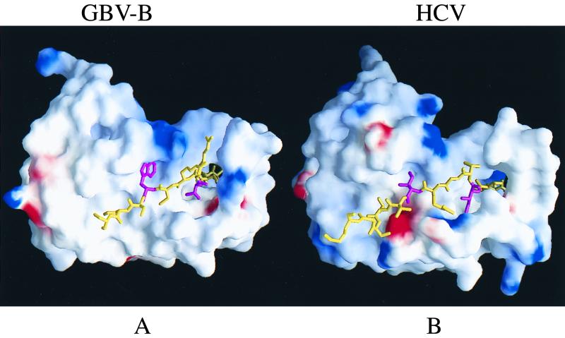
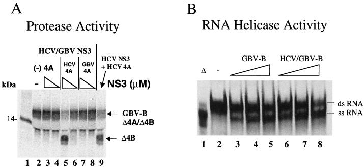
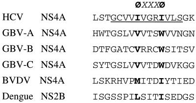
GB virus B (GBV-B) is closely related to hepatitis C virus (HCV) and causes acute hepatitis in tamarins (Saguinus species), making it an attractive surrogate virus for in vivo testing of anti-HCV inhibitors in a small monkey model. It has been reported that the nonstructural protein 3 (NS3) serine protease of GBV-B shares similar substrate specificity with its counterpart in HCV. Authentic proteolytic processing of the HCV polyprotein junctions (NS4A/4B, NS4B/5A, and NS5A/5B) can be accomplished by the GBV-B NS3 protease in an HCV NS4A cofactor-independent fashion. We further characterized the protease activity of a full-length GBV-B NS3 protein and its cofactor requirement using in vitro-translated GBV-B substrates. Cleavages at the NS4A/4B and NS5A/5B junctions were readily detectable only in the presence of a cofactor peptide derived from the central region of GBV-B NS4A. Interestingly, the GBV-B substrates could also be cleaved by the HCV NS3 protease in an HCV NS4A cofactor-dependent manner, supporting the notion that HCV and GBV-B share similar NS3 protease specificity while retaining a virus-specific cofactor requirement. This finding of a strict virus-specific cofactor requirement is consistent with the lack of sequence homology in the NS4A cofactor regions of HCV and GBV-B. The minimum cofactor region that supported GBV-B protease activity was mapped to a central region of GBV-B NS4A (between amino acids Phe22 and Val36) which overlapped with the cofactor region of HCV. Alanine substitution analysis demonstrated that two amino acids, Val27 and Trp31, were essential for the cofactor activity, a finding reminiscent of the two critical residues in the HCV NS4A cofactor, Ile25 and Ile29. A model for the GBV-B NS3 protease domain and NS4A cofactor complex revealed that GBV-B might have developed a similar structural strategy in the activation and regulation of its NS3 protease activity. Finally, a chimeric HCV/GBV-B bifunctional NS3, consisting of an N-terminal HCV protease domain and a C-terminal GBV-B RNA helicase domain, was engineered. Both enzymatic activities were retained by the chimeric protein, which could lead to the development of a chimeric GBV-B virus that depends on HCV protease function.


Figures

FIG. 1
FIG. 1
Schematic illustration of various plasmid constructs and cofactor peptides used in this study. The numbers above or below each construct or peptide indicate the amino acid positions corresponding to the published full-length polyprotein for each virus. The predicted molecular masses for each substrate and its cleavage products were indicated under each substrate plasmid.
FIG. 2
FIG. 2
Cofactor-dependent trans cleavage activity of GBV-B NS3 protease on HCV substrates. 35S-labeled in vitro-translated substrates from HCV cleavage sites were translated as described in Materials and Methods: NS4A/NS4B (Δ4A/Δ4B) (A), NS4B/NS5A (Δ4B/Δ5A) (B), and NS5A/NS5B (Δ5A/Δ5B) (C). Labeled substrates and purified GBV-B protease were incubated in the presence or absence of 20 μM GBV-B NS4A peptide (aa 16 to 46, Δ4A31) for 1.5 h at 30°C. Final concentrations of GBV-B protease were 250 nM for NS4A/4B and 5 μM for NS4B/5A and NS5A/5B. In parallel, cleavages of HCV substrates by purified HCV NS3/4A are shown in each panel (lane 2). Samples were analyzed on an SDS–15% PAGE gel and detected by phosphorimaging scan.
FIG. 3
FIG. 3
Cofactor-dependent trans cleavage activity of GBV-B NS3 protease on GBV-B substrates. 35S-labeled in vitro-translated substrates from GBV-B cleavage sites were mixed with 2 μM purified protease in the presence or absence of 20 μM GBV-B NS4A peptide (Δ4A31): Δ4A/Δ4B (A), Δ4B/Δ5A (B), and Δ5A/Δ5B (C). A mutant (NS3S137A) GBV-B protease was also tested (lane 4 of each panel). Proteins were separated and analyzed as described in the legend to Fig. 2.
FIG. 4
FIG. 4
Cofactor-dependent trans cleavage activity of HCV NS3 protease on GBV-B substrates. (A) trans cleavage reaction using GBV-B NS4A/4B as the substrate. Purified HCV full-length NS3 protease was mixed with translated substrate in the presence or absence of 100 μM HCV NS4A cofactor peptide (Δ4A13). Decreasing concentrations of protease were tested in this assay (indicated by a triangular descending slope: 2.5, 0.25, 0.025, and 0.0025 μM). (B) trans cleavage reaction using GBV-B NS 5A/5B as the substrate. HCV NS3 protease at 10 μM was tested in the presence or absence of 100 μM NS4A cofactor peptide (Δ4A13).
FIG. 5
FIG. 5
Mapping the minimum region within GBV-B NS4A required for the cofactor activity. Sequences of the entire HCV and GBV-B NS4A proteins are aligned at the top with the minimum cofactor region for HCV NS4A underlined. Vertical lines represent identity while colons represent similarity between the two viruses within the NS4A region. Dashes represent a gap created to maximize the homology alignment. To compare the cofactor activity, 35S-labeled GBV-B NS4A/4B substrate (Δ4A/Δ4B) was incubated with 1.5 μM GBV-B protease in the presence of various GBV-B NS4A peptides as shown in the figure. Cleavage products were then analyzed and detected by phosphorimaging and quantified by ImageQuant software (Molecular Dynamics). The cofactor activity was graded by comparing the enhancement of the protease activity to that of the background (cleavage activity in the absence of NS4A cofactor and scored as follows: +++, 11- to 20-fold; ++, 5- to 10-fold; +, 2.5- to 5-fold; ±, 1.5- to 2-fold; and −, ≤1.5-fold of the background activity. Amino acids critical for cofactor activation of the NS3 protease are shown in boldface, larger type; the minimum GBV-B NS4A cofactor region is also underlined.
FIG. 6
FIG. 6
NS4A cofactor activity is virus specific. HCV or GBV-B NS3 protease was tested in the absence of NS4A peptide or in the presence of either HCV NS4A (Δ4A13) or GBV-B NS4A (Δ4A17) at 100 μM. The assay was performed using the NS4A/4B substrates from either HCV (A) or GBV-B (B). HCV NS3 was tested at 50 nM for both substrates; GBV-B protease was tested at 250 nM for HCV substrate and at 2 μM for GBV-B substrate.
FIG. 7
FIG. 7
Cofactor activity of alanine-substituted GBV-B NS4A peptides (Δ4A17). (A) Sequence of alanine-substituted peptides used in this study, with the alanine residue shown in boldface. The A24 wild type [A24(WT)] is the same as the wild-type peptide. (B) Cofactor activity tested in the trans cleavage assay. A 200 μM concentration of each peptide was used in each reaction containing the GBV-B NS4A/4B substrate and 1.5 μM GBV-B NS3 protease. Cleavage products were analyzed similarly as described in the previous figures. The alanine-substituted residue and the corresponding amino acid position (number) in NS4A are shown above each lane. Vertical arrows identify the amino acids critical for cofactor activity. (C) Quantitative analysis of the cofactor activities (shown in panel B) expressed as the percent inhibition of wild-type cofactor activity.
FIG. 8
FIG. 8
Surface representation of GBV-B and HCV protease complexed with the corresponding 4A cofactors (shown as sticks). (A) GBV-B NS3/4A model. (B) HCV NS3/4A model. The electrostatic potential distribution is color coded: blue, positive; red, negative; white, neutral. The N-terminal 11 aa were removed to expose the detail interaction between NS4A and NS3 protease. The two critical Trp31 and Val27 (shown as magenta sticks in contrast to the rest of 4A colored yellow) reveal their location and complementary with the corresponding hydrophobic pockets, which is strikingly similar to that of HCV NS3/4A complex structure in panel B. The two essential residues Ile29 and Ile25, in HCV NS4A are also colored in magenta.
FIG. 9
FIG. 9
HCV protease and GBV-B RNA helicase activities of the chimeric NS3 protein. (A) Protease activity. The GBV-B NS4A/4B (Δ4A/4B) substrate was mixed with the chimeric HCV/GBV-B NS3 at 2 μM (lanes 3, 5, and 7) or at 0.07 μM (lanes 4, 6, and 8) in the absence NS4A cofactor or the presence of either HCV NS4A cofactor (Δ4A13) or GBV-B NS4A cofactor (Δ4A17). The final concentrations of the cofactor peptides were 100 μM. The cleavage products were analyzed as described previously. (B) RNA helicase activity. The RNA helicase activity of the chimeric NS3 was compared to that of the native GBV-B NS3. The Δ symbol indicates that the sample was boiled before loading onto the gel. Increasing amounts (2, 5, and 10 pmol) of each protein were tested for the dsRNA unwinding activity. The released ssRNAs were separated and analyzed as described in Materials and Methods. The ascending triangular slopes indicate the increasing concentration of the NS3 proteins. The “−” symbol indicates the no-enzyme controls.
FIG. 10
FIG. 10
Cofactor activation motif for the flavivirus-like NS3 serine protease. Cofactor regions from various members of the Flaviviridae family are compared. The minimum cofactor region in HCV is underlined. A common feature involving two bulky hydrophobic amino acids (in boldface type symbolized by a “Φ”) is proposed to form the activation motif, ΦxxxΦ, where “x” is any amino acid.

Similar articles

Cited by

References

    1. Alter H J, Purcell R H, Shih J W, Melpolder J C, Houghton M, Choo Q-L, Kuo G. Detection of antibody to hepatitis C virus in prospectively followed transfusion recipients with acute and chronic non-A non-B hepatitis. N Engl J Med. 1989;321:1494–1500. - PubMed
    1. Alter M J, Kruszon-Moran D, Nainan O V, McQuillan G M, Gao F, Moyer L A, Kaslow R A, Margolis H S. The prevalence of hepatitis C virus infection in the United States, 1988 through 1994. N Engl J Med. 1999;341:556–562. - PubMed
    1. Bartenschlager R, Ahlborn-Laake L, Mous J, Jacobsen H. Nonstructural protein 3 of the hepatitis C virus encodes a serine-type proteinase required for cleavage at the NS3/4 and NS4/5 junctions. J Virol. 1993;67:3835–3844. - PMC - PubMed
    1. Beard M R, Abell G, Honda M, Carroll A, Gartland M, Clarke B, Suzuki K, Lanford R, Sangar D V, Lemon S M. An infectious molecular clone of a Japanese genotype 1b hepatitis C virus. Hepatology. 1999;30:316–624. - PubMed
    1. Behrens S-E, Tomei L, De Francesco R. Identification and properties of the RNA-dependent RNA polymerase of hepatitis C virus. EMBO J. 1996;15:12–22. - PMC - PubMed

Publication types

MeSH terms

LinkOut - more resources