. 2000 May;74(9):4310-8.
doi: 10.1128/jvi.74.9.4310-4318.2000.

Brome mosaic virus polymerase-like protein 2a is directed to the endoplasmic reticulum by helicase-like viral protein 1a

Affiliations

Brome mosaic virus polymerase-like protein 2a is directed to the endoplasmic reticulum by helicase-like viral protein 1a

J Chen et al. J Virol. 2000 May.

Abstract

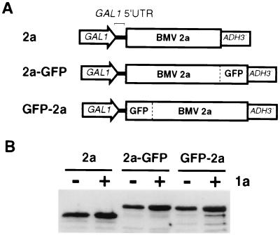
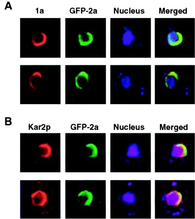
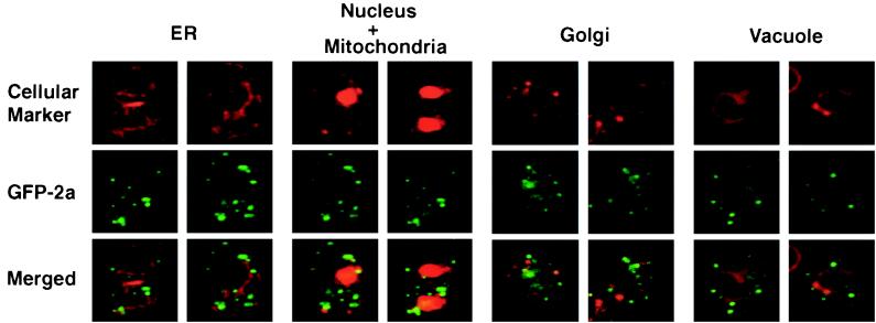
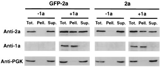
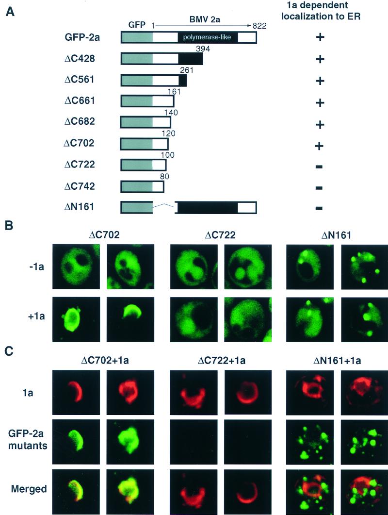
Brome mosaic virus (BMV), a positive-strand RNA virus in the alphavirus-like superfamily, encodes RNA replication proteins 1a and 2a. 1a contains a C-terminal helicase-like domain and an N-terminal domain implicated in viral RNA capping, and 2a contains a central polymerase-like domain. 1a and 2a colocalize in an endoplasmic reticulum (ER)-associated replication complex that is the site of BMV-specific RNA-dependent RNA synthesis in plant and yeast cells. 1a also localizes to the ER in the absence of 2a or viral RNA replication templates. To investigate the determinants of 2a localization, we fused 2a to the green fluorescent protein (GFP), creating a functional GFP-2a fusion that supported BMV RNA replication and subgenomic mRNA transcription. In the absence of 1a, the GFP-2a fusion was found to be diffused throughout the cytoplasm and in punctate spots not associated with any cytoplasmic organelle so far tested. Formation of these spots was dependent on the C-terminal half of 2a and may represent aggregation of a fraction of 2a. When coexpressed with 1a, GFP-2a colocalized with 1a and ER-resident protein Kar2p in a partial or complete ring around the nucleus. Consistent with these results, cell fractionation showed that both the GFP-2a fusion and wild-type (wt) 2a remained soluble when expressed alone, while in cells coexpressing 1a, most of the GFP-2a fusion or wt 2a cofractionated with 1a in the rapidly sedimenting membrane fraction. Deletion analysis showed that the N-terminal 120-amino-acid segment of 2a, containing one of two 2a regions previously shown to interact with 1a, was necessary and sufficient for 1a-directed localization of GFP-2a derivatives to the ER. These results suggest that 1a, which also interacts independently with the ER and viral RNA, is a key organizer of RNA replication complex assembly.


Figures

FIG. 1
FIG. 1
Expression of BMV 2a and GFP-fused 2a derivatives in yeast. (A) Schematic representation of expression cassettes for 2a and the fusions. The galactose-inducible GAL1 promoter, GAL1 5′ untranslatable region (5′ UTR), 2a- and GFP-coding sequences, and ADH1 polyadenylation signal are indicated. The expression cassettes were assembled into the multiple-cloning sites of Ycplac111, a yeast CEN4 centromeric plasmid, for protein expression in yeast. (B) Immunoblot analysis of 2a and the fusions. Yeast cells were transformed with plasmids expressing wt 2a or the fusions either alone (−1a) or together with a plasmid expressing 1a (+1a). Cells were grown in galactose medium to induce protein expression and harvested at mid-log phase. Total proteins were extracted and subjected to 0.1% SDS–10% PAGE and immunoblot analysis with anti-2a monoclonal antibodies.
FIG. 2
FIG. 2
GFP-fused 2a derivatives support BMV RNA3 replication and transcription. 1a- and RNA3-expressing plasmids were cotransformed into yeast with plasmids expressing either wt 2a, the indicated fusion, or the starting plasmid lacking 2a sequences (−2a) as indicated. Equal amounts of total RNA prepared from the resulting galactose-induced yeast were analyzed by Northern blotting with a single-stranded, 32P-labeled RNA probe complementary to either positive- or negative-strand RNA3 as indicated. (A) Representative Northern blots with the migration positions of each virion RNA indicated. (B) Average relative accumulation and standard deviation of RNA3 and RNA4 replication products, as determined for three independent transformants of each 2a derivative. Values are shown as percentage of RNA3 or RNA4 accumulation in yeast cells expressing wt 2a.
FIG. 3
FIG. 3
Colocalization of GFP-2a with 1a and perinuclear ER in yeast coexpressing GFP-2a, 1a, and RNA3. Yeast cotransformed with plasmids expressing GFP-2a, 1a, and RNA3 was grown in galactose medium to induce protein expression and harvested in mid-log phase. Cells were then fixed with formaldehyde, treated with Lyticase to remove the cell wall, and incubated with polyclonal antibodies against 1a (A) or ER-resident protein Kar2p (B) and finally treated with Texas red-labelled secondary antibodies. After secondary antibody treatment, cells were incubated briefly with the DNA stain TO-PRO-3 iodide to visualize the nucleus. For each cell, the differentially fluorescing protein (red, 1a or Kar2p; green, GFP-2a) and DNA (blue) images were gathered simultaneously from the same optical section with a multichannel confocal microscope and appropriate filters. Control experiments omitting 1a or Kar2p antibodies, DNA stain, or GFP-2a confirmed that there was no signal leakage from any channel into the other channels. The three images were digitally superimposed (Merged) to depict the relationship among GFP-2a, 1a, ER, and nucleus. Two representative images of individual yeast cells are shown for each case. Each image measures 7 μm per side.
FIG. 4
FIG. 4
1a-induced redistribution of GFP-2a to perinuclear ER in yeast. A plasmid expressing either wt 2a, free GFP, or GFP-2a was transformed into yeast either alone (−1a) or together with a plasmid expressing 1a (+1a). Cells were grown in galactose medium, harvested at mid-log phase, immobilized on glass slides with a thin layer of 1% agarose gel in synthetic galactose medium, and visualized on a confocal microscope by direct GFP fluorescence. Except for yeast expressing wt 2a, two representative images of individual yeast cells are shown for each case. Each image measures 7 μm per side. Arrowheads indicate spots of green fluorescence.
FIG. 5
FIG. 5
Relation of GFP-2a localization to cellular organelles in the absence of 1a. Yeast cells expressing GFP-2a alone were fixed with formaldehyde, treated with Lyticase to remove the cell wall, and incubated with antibodies against proteins localizing on the ER, Golgi apparatus, or vacuolar membranes, respectively. DNA stain TO-PRO-3 iodide was used to visualize the mitochondria and the nucleus. For each cell, the differentially fluorescing protein and DNA images were gathered simultaneously with appropriate filters as described in the legend to Fig. 3. The two images were digitally superimposed (Merged) to depict the distribution of GFP-2a relative to cellular organelles. Two representative images of individual yeast cells are shown for each case. Each image measures 7 μm per side.
FIG. 6
FIG. 6
Effects of 1a on GFP-2a and wt 2a distribution by cell fractionation. Yeast cells expressing either GFP-2a or wt 2a alone (−1a) or coexpressing 1a (+1a) were grown in galactose medium and harvested at mid-log phase. The cells were then treated with Lyticase to remove the cell wall, and the resulting spheroplasts were lysed osmotically to yield a total protein fraction (Tot.). A portion of the lysate was then subjected to low-speed centrifugation to yield pellet (Pell.) and supernatant (Sup.) fractions. Equal percentages of each fraction were subjected to 0.1% SDS–10% PAGE and immunoblot analysis with antibodies against 2a, 1a, or PGK, a cytoplasmic soluble protein.
FIG. 7
FIG. 7
The N-terminal 120 residues of 2a are required and sufficient for 1a-directed localization of GFP-2a to the ER. (A) Schematic representation of full-length GFP-2a and 2a C-terminal truncations and a 2a N-terminal in-frame deletion derived from it. GFP and 2a segments and the conserved polymerase-like domain of 2a are indicated. + and − indicate the ability of each construct to be directed to perinuclear ER by 1a as determined by direct fluorescence confocal microscopy of live yeast. (B) Representative images of the intracellular distribution of the indicated GFP-2a derivatives in live yeast either coexpressing 1a (+1a) or lacking 1a (−1a). Direct fluorescence confocal microscopy was performed as described in the legend to Fig. 4. (C) Localization of 1a and GFP-2a derivatives in yeast coexpressing 1a and the indicated GFP-2a derivatives. 1a was immunostained as described in the legend to Fig. 3. Two representative images of individual yeast cells are shown for each case. Each image measures 7 μm per side.

References

    1. Ahlquist P. Bromovirus RNA replication and transcription. Curr Opin Genet Dev. 1992;2:71–76. - PubMed
    1. Ahola T, Ahlquist P. Putative RNA capping activities encoded by brome mosaic virus: methylation and covalent binding of guanylate by replicase protein 1a. J Virol. 1999;73:10061–10069. - PMC - PubMed
    1. Allison R, Thompson C, Ahlquist P. Regeneration of a functional RNA virus genome by recombination between deletion mutants and requirement for cowpea chlorotic mottle virus 3a and coat genes for systemic infection. Proc Natl Acad Sci USA. 1990;87:1820–1824. - PMC - PubMed
    1. Argos P. A sequence motif in many polymerases. Nucleic Acids Res. 1988;16:9909–9916. - PMC - PubMed
    1. Ausubel F M, Brent R, Kingston R E, Moore D D, Seidman J G, Smith J A, Struhl K, editors. Current protocols in molecular biology. New York, N.Y: John Wiley & Sons; 1987.

Publication types

MeSH terms

LinkOut - more resources