. 2000 May;74(9):4319-26.
doi: 10.1128/jvi.74.9.4319-4326.2000.

Infectious bronchitis virus E protein is targeted to the Golgi complex and directs release of virus-like particles

Affiliations

Infectious bronchitis virus E protein is targeted to the Golgi complex and directs release of virus-like particles

E Corse et al. J Virol. 2000 May.

Abstract

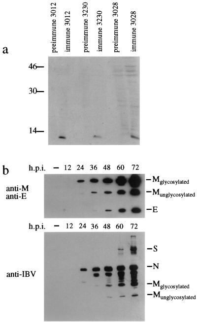
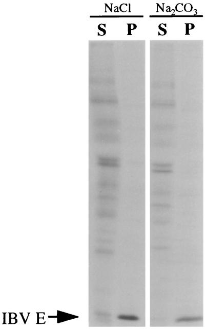
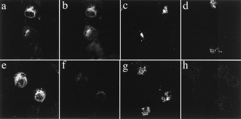
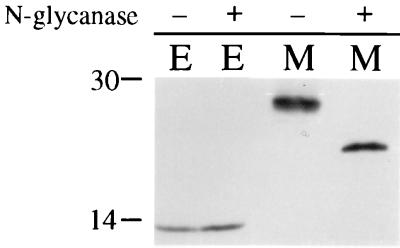
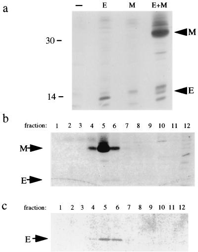
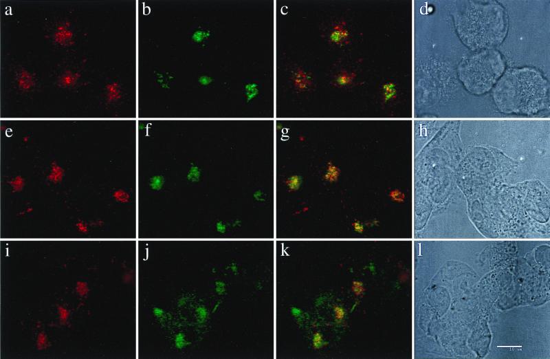
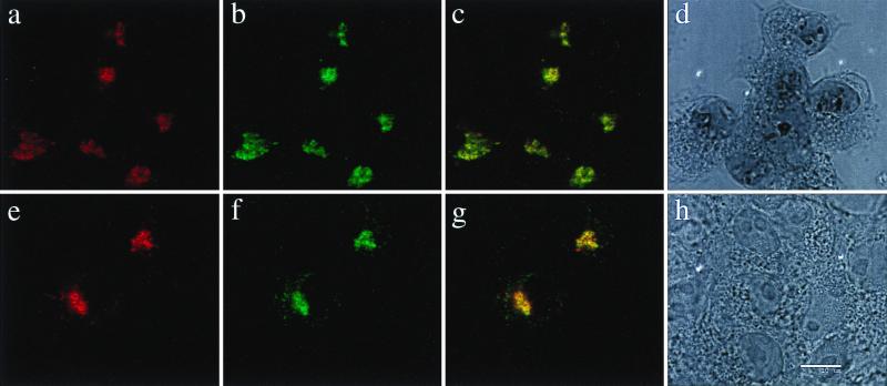
The coronavirus E protein is a poorly characterized small envelope protein present in low levels in virions. We are interested in the role of E in the intracellular targeting of infectious bronchitis virus (IBV) membrane proteins. We generated a cDNA clone of IBV E and antibodies to the E protein to study its cell biological properties in the absence of virus infection. We show that IBV E is an integral membrane protein when expressed in cells from cDNA. Epitope-specific antibodies revealed that the C terminus of IBV E is cytoplasmic and the N terminus is translocated. The short luminal N terminus of IBV E contains a consensus site for N-linked glycosylation, but the site is not used. When expressed using recombinant vaccinia virus, the IBV E protein is released from cells at low levels in sedimentable particles that have a density similar to that of coronavirus virions. The IBV M protein is incorporated into these particles when present. Indirect immunofluorescence microscopy showed that E is localized to the Golgi complex in cells transiently expressing IBV E. When coexpressed with IBV M, both from cDNA and in IBV infection, the two proteins are colocalized in Golgi membranes, near the coronavirus budding site. Thus, even though IBV E is present at low levels in virions, it is apparently expressed at high levels in infected cells near the site of virus assembly.


Figures

FIG. 1
FIG. 1
Antibodies recognizing the IBV E protein are specific. (a) BHK-21 cells infected with vvIBVE were radiolabeled from 3.5 to 4.5 h postinfection and lysed in detergent solution. The lysates were immunoprecipitated with polyclonal peptide antibodies to IBV E and the corresponding preimmune sera. The immunoprecipitates were analyzed by SDS-PAGE on a 15% polyacrylamide gel and visualized by fluorography. 3012 is the rabbit anti-C-terminal peptide antibody, 3230 is the rat anti-C-terminal peptide antibody, and 3028 is the rabbit anti-N-terminal peptide antibody. (b) Vero cells infected with IBV were harvested after infection for the times shown in SDS sample buffer. The samples were subjected to SDS-PAGE and Western blotting with anti-E and anti-M antibodies (top) or anti-IBV antibodies (bottom). The lanes labeled − contain lysate from mock-infected cells. h.p.i., hours postinfection.
FIG. 2
FIG. 2
IBV E is an integral membrane protein. BHK-21 cells infected with vvIBVE were radiolabeled from 3.5 to 4.5 h postinfection, and microsomes were prepared from homogenized cells. The microsomes were extracted with either 0.1 M NaCl (left) or 0.1 M Na2CO3, (pH 11.5) (right), and the membranes were pelleted. Pellets (P) and supernatants (S) were immunoprecipitated with anti-IBV E in the presence of detergent, and the immunoprecipitates were analyzed by SDS-PAGE on a 15% polyacrylamide gel and visualized by fluorography.
FIG. 3
FIG. 3
The C terminus of IBV E is cytoplasmic. BHK-21 cells were infected with vTF7-3 and transfected with a plasmid encoding the Golgi resident protein Gm1 (a, b, e, and f) or infected with vvIBV E (c, d, g, and h) as described in Materials and Methods. At 6 h postinfection, cells were either permeabilized with digitonin (e to h) and fixed for immunofluorescence or fixed and permeabilized with Triton X-100 prior to staining (a to d). The cells were stained with antibodies to the cytoplasmic tail of Gm1 (a and e), the luminal head of Gm1 (b and f), the C terminus of IBV E (c and g), or the N terminus of IBV E (d and h). Secondary antibodies were fluorescein-conjugated goat anti-mouse IgG (b and f) and Texas red-conjugated goat anti-rabbit IgG (all other panels).
FIG. 4
FIG. 4
The N-terminal N-linked glycosylation site of IBV E is not used. BHK-21 cells infected with vTF7-3 were transfected with plasmids encoding IBV E or IBV M, radiolabeled from 3.5 to 4.5 h postinfection and lysed in detergent solution. The lysates were immunoprecipitated with appropriate antibodies to E or M. The immunoprecipitates were mock treated (−) or treated with N-glycanase (+), analyzed by SDS-PAGE on a 15% polyacrylamide gel, and visualized by fluorography.
FIG. 5
FIG. 5
IBV E is released from transfected cells in VLPs that incorporate IBV M if present. (a) BHK-21 cells were mock infected (−), infected with vvIBVE (E), vvIBVM (M), or vvIBVE plus vvIBVM (E+M), radiolabeled from 3.5 to 4.5 h postinfection, and chased for 3 h. Medium was collected, cleared of debris, and immunoprecipitated with anti-E and anti-M antibodies. The immunoprecipitates were analyzed by SDS-PAGE and visualized by fluorography. (b) BHK-21 cells coinfected with vvIBVE and vvIBVM were radiolabeled from 3.5 to 4.5 h postinfection and chased for 3 h. Concentrated supernatants were loaded onto a continuous 20 to 55% sucrose gradient and centrifuged to equilibrium. Fractions were collected and immunoprecipitated with anti-E and anti-M antibodies. The immunoprecipitates were separated by SDS-PAGE and visualized by fluorography. Fraction 1 corresponds to the top of the gradient, and fraction 12 corresponds to the bottom of the gradient. (c) BHK-21 cells were infected with vvIBVE only and treated exactly as described for panel b.
FIG. 6
FIG. 6
The IBV E protein is localized to the Golgi complex. BHK-21 cells infected with vvIBVE were fixed for immunofluorescence at 6 h postinfection, permeabilized with Triton X-100, and double labeled with antibodies to IBV E and different endogenous Golgi resident proteins. (a to d) Cells were stained with rabbit anti-IBV E and mouse anti-GM130; (e to h) cells were stained with rabbit anti-IBV E and mouse anti-mannosidase II; (i to l) cells were stained with rabbit anti-IBV E and mouse anti-syntaxin 6. Secondary antibodies were Texas red-conjugated goat anti-rabbit IgG and fluorescein-conjugated goat anti-mouse IgG. In each row, the red image corresponds to IBV E staining and the green image corresponds to the appropriate Golgi marker. The third image in each row (c, g, and k) is a merged image, where yellow represents overlap between the red- and green-staining patterns. The fourth image in each row (d, h, and l) is a phase image of each field of labeled cells. Bar, 10 μm.
FIG. 7
FIG. 7
IBV E and IBV M colocalize in transfected and IBV-infected cells. BHK-21 cells infected with vvIBVE and vvIBVM (a to d) or IBV-infected Vero cells (e to h) were fixed for immunofluorescence at 6 h postinfection and double labeled with rat anti-E antibody and rabbit anti-M antibody. Secondary antibodies were Texas red-conjugated goat anti-rat IgG and fluorescein-conjugated goat anti-rabbit IgG. The red images correspond to IBV E staining, and the green images correspond to IBV M staining. The third image in each row (c and g) is a merged image, where yellow represents overlap between the red- and green-staining patterns. The fourth image in each row (d and h) is a phase image of the field of labeled cells. Bar, 10 μm.

References

    1. Abraham S, Kienzle T E, Lapps W E, Brian D A. Sequence and expression analysis of potential nonstructural proteins of 4.9, 4.8, 12.7, and 9.5 kDa encoded between the spike and membrane protein genes of the bovine coronavirus. Virology. 1990;177:488–495. - PMC - PubMed
    1. An S, Chen C J, Leibowitz J L, Makino S. Induction of apoptosis in murine coronavirus-infected cultured cells and demonstration of E protein as an apoptosis inducer. J Virol. 1999;73:7853–7859. - PMC - PubMed
    1. Baudoux P, Carrat C, Besnardeau L, Charley B, Laude H. Coronavirus pseudoparticles formed with recombinant M and E proteins induce alpha interferon synthesis by leukocytes. J Virol. 1998;72:8636–8643. - PMC - PubMed
    1. Bos E C W, Luytjes W, Van der Meulen H, Koerten H K, Spaan W J M. The production of recombinant infectious DI-particles of a murine coronavirus in the absence of helper virus. Virology. 1996;218:52–60. - PMC - PubMed
    1. Bock J B, Klumperman J, Davanger S, Scheller R H. Syntaxin 6 functions in trans-Golgi network vesicle trafficking. Mol Biol Cell. 1997;8:1261–1271. - PMC - PubMed

Publication types

MeSH terms

Substances

LinkOut - more resources