. 2000 May;74(9):4361-76.
doi: 10.1128/jvi.74.9.4361-4376.2000.

Unusual polymorphisms in human immunodeficiency virus type 1 associated with nonprogressive infection

Affiliations

Unusual polymorphisms in human immunodeficiency virus type 1 associated with nonprogressive infection

L Alexander et al. J Virol. 2000 May.

Abstract

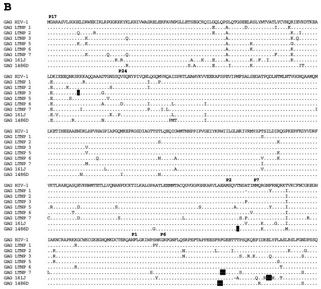
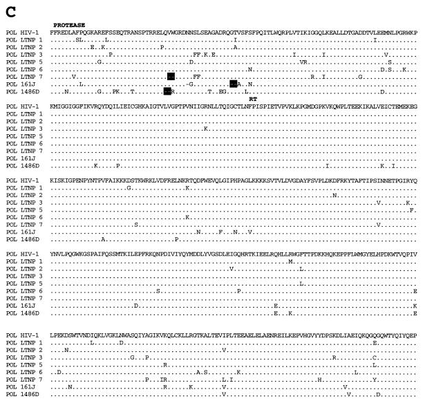
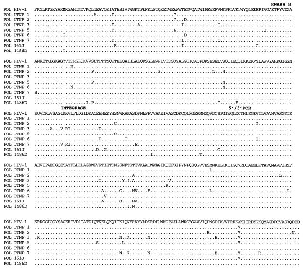
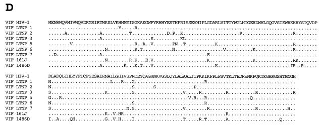
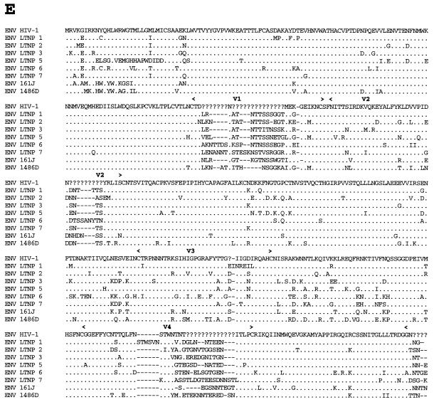
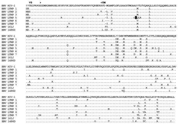
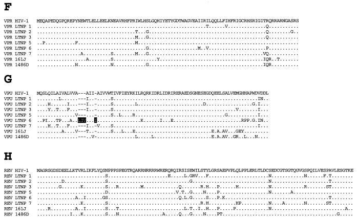
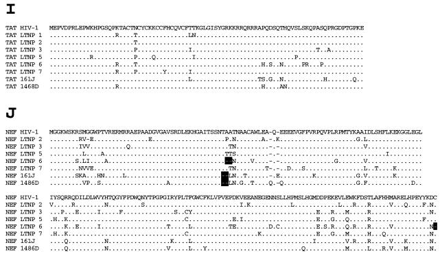
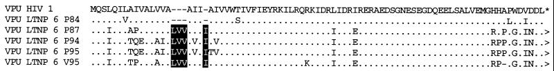
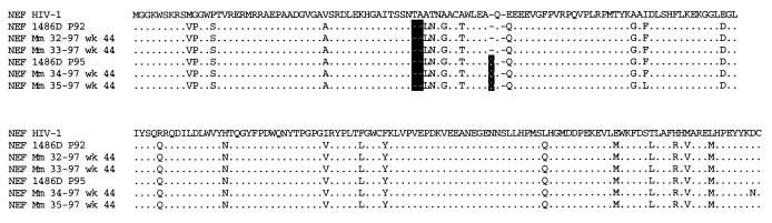
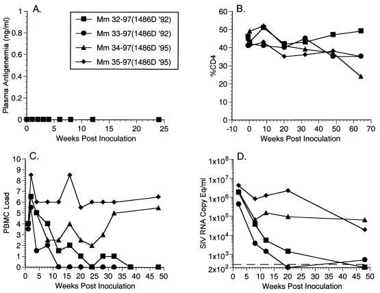
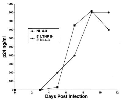
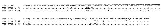
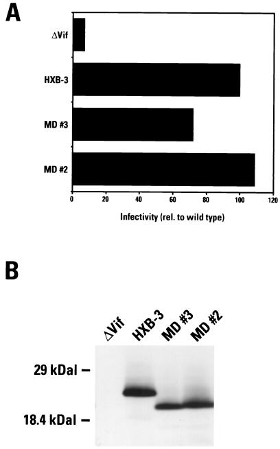
Factors accounting for long-term nonprogression may include infection with an attenuated strain of human immunodeficiency virus type 1 (HIV-1), genetic polymorphisms in the host, and virus-specific immune responses. In this study, we examined eight individuals with nonprogressing or slowly progressing HIV-1 infection, none of whom were homozygous for host-specific polymorphisms (CCR5-Delta32, CCR2-64I, and SDF-1-3'A) which have been associated with slower disease progression. HIV-1 was recovered from seven of the eight, and recovered virus was used for sequencing the full-length HIV-1 genome; full-length HIV-1 genome sequences from the eighth were determined following amplification of viral sequences directly from peripheral blood mononuclear cells (PBMC). Longitudinal studies of one individual with HIV-1 that consistently exhibited a slow/low growth phenotype revealed a single amino acid deletion in a conserved region of the gp41 transmembrane protein that was not seen in any of 131 envelope sequences in the Los Alamos HIV-1 sequence database. Genetic analysis also revealed that five of the eight individuals harbored HIV-1 with unusual 1- or 2-amino-acid deletions in the Gag sequence compared to subgroup B Gag consensus sequences. These deletions in Gag have either never been observed previously or are extremely rare in the database. Three individuals had deletions in Nef, and one had a 4-amino-acid insertion in Vpu. The unusual polymorphisms in Gag, Env, and Nef described here were also found in stored PBMC samples taken 3 to 11 years prior to, or in one case 4 years subsequent to, the time of sampling for the original sequencing. In all, seven of the eight individuals exhibited one or more unusual polymorphisms; a total of 13 unusual polymorphisms were documented in these seven individuals. These polymorphisms may have been present from the time of initial infection or may have appeared in response to immune surveillance or other selective pressures. Our results indicate that unusual, difficult-to-revert polymorphisms in HIV-1 can be found associated with slow progression or nonprogression in a majority of such cases.


Figures

FIG. 1
FIG. 1
Subtyping of the HIV-1 sequence of LTNP 1486D. 1486D sequences from bp 297 to 9607 (numbers based on NL 4-3 sequences) were analyzed using the Los Alamos subtyping program and subtyping reference sequences. The score value indicates the relative relatedness of 1486D sequences at different loci throughout the genome. The color of the bar at the top indicates the subtype that 1486D sequences are most closely related to at loci throughout the HIV-1 genome.
FIG. 2
FIG. 2
Dendrograms of reference sequences of HIV-1 isolates of different subtypes and LTNP sequences for Gag (A) and Nef (B). These analyses were performed using PAUP. Bootstrap confidence values were assigned when a minimum value of 70 was achieved for a particular branch. The dendrogram was rooted to the SIVcpzGAB sequences. The identification of the subtype isolates which are symbolized here is described in Material and Methods.
FIG. 3
FIG. 3
Alignments of LTNP sequences. Nucleotide (A) and amino acid (B to J) sequences of the indicated HIV-1 genetic elements and proteins were aligned to the group M, subgroup B, consensus sequences (HIV-1). Periods indicate conservation between LTNP and consensus B sequences. A dash indicates that a base or amino acid is not contained in a particular sequence. Unusual polymorphisms consistently observed in HIV-1 sequences from LTNPs are highlighted in black. The data presented represent consensus sequences from two clones from each of two independent PCRs.
FIG. 3
FIG. 3
Alignments of LTNP sequences. Nucleotide (A) and amino acid (B to J) sequences of the indicated HIV-1 genetic elements and proteins were aligned to the group M, subgroup B, consensus sequences (HIV-1). Periods indicate conservation between LTNP and consensus B sequences. A dash indicates that a base or amino acid is not contained in a particular sequence. Unusual polymorphisms consistently observed in HIV-1 sequences from LTNPs are highlighted in black. The data presented represent consensus sequences from two clones from each of two independent PCRs.
FIG. 4
FIG. 4
Alignment of LTNP 6 Vpu sequences. LTNP 6 amino acid sequences from the time points indicated (e.g., P84, PBMC sample obtained in 1984) were aligned to the group M, subgroup B, consensus sequences (HIV-1). V, sequences were obtained from isolated HIV-1. Each sequence represents a consensus of two clones from each of two independent PCRs. Periods indicate conservation between LTNP 6 and the consensus B sequences. A dash indicates that an amino acid is not contained in a particular sequence. The 4-aa insertion observed in Vpu sequences from LTNP 6 is highlighted in black.
FIG. 5
FIG. 5
Amino acid sequences of 1486D Nef after infection into rhesus monkeys with SHIVnef recombinants. A SHIVnef recombinant containing 1486D Nef sequences from PBMC obtained in 1992 (P92) was inoculated into Mm 32-97 and Mm 33-97, and a recombinant containing 1486D Nef sequences obtained in 1995 was inoculated into Mm 34-97 and Mm 35-97. The sequences in the inoculum and sequences obtained from the infected animal 44 weeks postinoculation were aligned to the group M, subgroup B, consensus (HIV-1) sequences. Each sequence represents a consensus of two clones from each of two independent PCRs. Periods indicate conservation between 1486D and the consensus B sequences. A dash indicates that an amino acid is not contained in a particular sequence. The deletion and insertion polymorphisms observed in Nef sequences from 1486D are highlighted in black.
FIG. 6
FIG. 6
LTNP p6gag-mediated Vpr incorporation. Recombinant HIV-1 containing LTNP p6gag sequences were purified by centrifugation. Virally associated proteins were separated by SDS-PAGE and electroblotted onto a membrane filter. HIV-1 Vpr was detected using a Vpr-specific polyclonal antiserum. Lane 1, NL 4-3 parental virus; lane 2, NL 4-3ΔVpr; lanes 3 to 10, recombinant HIV-1 containing LTNP p6gag sequences: lane 3, LTNP 2; lane 4, LTNP 1; lane 5, LTNP 3; lane 6, LTNP 5; lane 7, LTNP 6; lane 8, LTNP 7; lane 9, 161J; lane 10, 1486D.
FIG. 7
FIG. 7
Plasma antigenemia, CD4 percentage, PBMC load, and RNA copy (equivalents per milliliter) measurements for animals infected with SHIVnef recombinants containing 1486D Nef sequences. (A) Plasma antigenemia in SHIVnef-infected rhesus monkeys. p27 concentrations in plasma were determined at the time points indicated. The limit of detection is approximately 0.05 ng/ml. The week 0 sample is a preinfection sample taken immediately before inoculation with SHIVnef. (B) CD4 percentages in SHIVnef-infected rhesus monkeys. Whole blood was drawn from SHIVnef-inoculated animals at various times postinoculation and stained with OKT4, a fluorescein isothiocyanate-conjugated murine monoclonal antibody that was raised against rhesus macaque CD4 (American Type Culture Collection). The stained samples were analyzed using a FACSscan flow cytometer (Becton Dickinson). (C) Frequencies of infectious cells in PBMC of SHIVnef-infected rhesus macaques. Viral loads were graded on a scale from 0 to 10 indicating the number of PBMC needed to recover SIV. A value of 0 denotes that no virus was recovered using 106 cells, 1 denotes successful virus recovery from 106 cells, and 2 to 10 denote successful virus recovery from 333,333, 111,111, 37,037, 12,345, 4,115, 1,371, 457, 152, or 51 cells, respectively. (D) Plasma SIV RNA levels at the indicated weeks postinoculation for animals infected with SHIVnef recombinants. The dashed line indicates the threshold sensitivity of the assay, 300 copy eq/ml. A value of 0 was assumed for week 0.
FIG. 8
FIG. 8
Growth of recombinant HIV-1 containing 5′ LTNP 5 sequences. LTNP 5 and NL 4-3 sequences from bp 297 to 4402 (5′) and NL 4-3 sequences from bp 4402 to 9607 (3′) were amplified separately. An XmaI (CCCGGG) site was introduced at the junction of the PCR fragments (bp 4402) as described in Materials and Methods. XmaI-digested 5′ LTNP 5 and 5′ NL 4-3 fragments were cotransfected with 3′ NL 4-3 fragments into permissive CEMx174 cells. Supernatants were collected and assayed for p27 antigen concentration.
FIG. 9
FIG. 9
Alignment of LTNP 5 Vif sequences. The product of LTNP 5 vif allele representing the consensus (allele 2) and the product of an allele representing the most divergent sequences from the group M, subgroup B, consensus (allele 3) were aligned to subgroup B consensus sequences (HIV-1). Each sequence represents a consensus of two clones from each of two independent PCRs. A period indicates conservation between subgroup B and LTNP 5 sequences.
FIG. 10
FIG. 10
Vif functional assay. (A) LTNP 5 (MD) vif sequences 2 and 3 as well as HXB-3 vif were cotransfected into H9 cells with a vif-deficient proviral vector. Harvested viruses were used to challenge the indicator cell line C8166/HIV-CAT. Levels of CAT activity indicate levels of Vif-mediated infectivity. These data represent the means of four independent experiments. (B) Whole-cell lysates of transfected H9 cells were separated by SDS-PAGE and electroblotted onto a membrane filter. HIV-1 Vif was detected using a Vif-specific monoclonal antibody.

Similar articles

Cited by

References

    1. Adachi A, Gendelman H E, Koenig S, Folks T, Willey R, Rabson A, Martin M. Production of acquired immunodeficiency syndrome-associated retrovirus in human and nonhuman cells transfected with an infectious molecular clone. J Virol. 1986;59:284–291. - PMC - PubMed
    1. Alexander L, Du Z, Howe A Y M, Czajak S, Desrosiers R C. Induction of AIDS in rhesus monkeys by a recombinant simian immunodeficiency virus expressing nef of human immunodeficiency virus type 1. J Virol. 1999;73:5814–5825. - PMC - PubMed
    1. Barnes W M. PCR amplification of up to 35-kb DNA with high fidelity and high yield from lambda bacteriophage templates. Proc Natl Acad Sci USA. 1994;91:2216–2220. - PMC - PubMed
    1. Bebenek K, Abbotts J, Roberts J D, Wilson S H, Kunkel T A. Specificity and mechanism of error-prone replication by human immunodeficiency virus-1 reverse transcriptase. J Biol Chem. 1989;264:16948–16956. - PubMed
    1. Binley J M, Jin X, Huang Y, Zhang L, Cao Y, Ho D D, Moore J P. Persistent antibody responses but declining cytotoxic T-lymphocyte responses to multiple human immunodeficiency virus type 1 antigens in a long-term nonprogressing individual with a defective p17 proviral sequence and no detectable viral RNA expression. J Virol. 1998;72:3472–3474. - PMC - PubMed

Publication types

MeSH terms