

Increased expression of osteonectin/SPARC mRNA and protein in age-related human cataracts and spatial expression in the normal human lens
- PMID: 10756178
- PMCID: PMC2831409
Increased expression of osteonectin/SPARC mRNA and protein in age-related human cataracts and spatial expression in the normal human lens
Abstract
Purpose: We have previously reported increased levels of Osteonectin/SPARC transcript in age-related cataractous compared to normal human lenses. The purpose of the present study was to evaluate the corresponding levels of osteonectin/SPARC protein in age-related cataractous relative to normal lenses and to evaluate the levels of osteonectin/SPARC transcript in specific types of age-related human cataracts. The spatial expression of osteonectin/SPARC was also evaluated in normal human lenses.
Methods: Specific types of age-related cataracts were collected and graded. Normal human lenses were microdissected into epithelia and fibers. Osteonectin/SPARC protein levels were monitored by Western immunoblotting, and transcript levels were evaluated by reverse transcriptase polymerase chain reaction (RT-PCR). Osteonectin/SPARC expression patterns were examined by RT-PCR and by immunostaining.
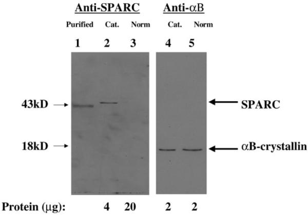
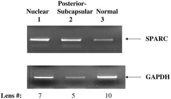
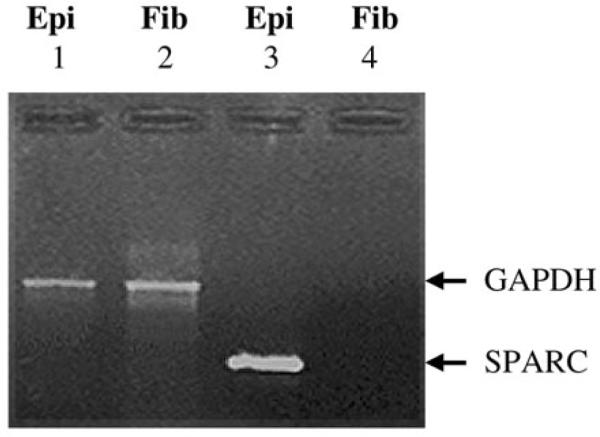
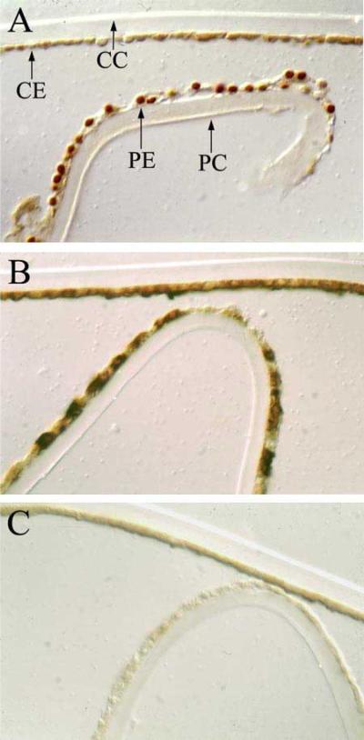
Results: Higher levels of osteonectin/SPARC protein were detected in age-related cataractous relative to normal human lenses. Increased levels of osteonectin/SPARC transcript were also detected in posterior-subcapsular and nuclear cataractous lenses relative to normal lenses. Osteonectin/SPARC transcripts were detected in the lens epithelium but not fibers. Osteonectin/SPARC protein levels were highest in the peripheral lens epithelium.
Conclusions: Consistent with our previous studies on osteonectin/SPARC mRNA levels, osteonectin/SPARC protein levels were also elevated in cataractous compared to normal human lenses. Increased levels of osteonectin/SPARC mRNA were also found in nuclear and posterior-subcapsular cataracts relative to normal lenses. Osteonectin/SPARC expression is confined to the lens epithelium, and osteonectin/SPARC levels are highest in the peripheral lens epithelium.
Figures
References
-
- Spector A. Aging of the lens and cataract formation. In: Sekuler R, Kline D, Dismukes K, editors. Aging and human visual function. A.R. Liss; New York: 1982. pp. 27–43.
-
- Brown NAP, Bron AJ. Lens disorders: a clinical manual of cataract diagnosis. Butterwoth-Heinemasnn; Oxford: 1996. pp. 53–133.
-
- Spector A. Oxidative stress-induced cataract: mechanism of action. FASEB J. 1995;9:1173–82. - PubMed
-
- Hightower KR. The role of the lens epithelium in development of UV cataract. Curr Eye Res. 1995;14:71–8. - PubMed
-
- Piatigorsky J. Lens differentiation in vertebrates. A review of cellular and molecular features. Differentiation. 1981;19:134–53. - PubMed
Publication types
MeSH terms
Substances
Grants and funding
LinkOut - more resources
Full Text Sources
Other Literature Sources
Medical
Miscellaneous
