. 2000 May;20(9):2959-69.
doi: 10.1128/MCB.20.9.2959-2969.2000.

Negative and translation termination-dependent positive control of FLI-1 protein synthesis by conserved overlapping 5' upstream open reading frames in Fli-1 mRNA

Affiliations

Negative and translation termination-dependent positive control of FLI-1 protein synthesis by conserved overlapping 5' upstream open reading frames in Fli-1 mRNA

S Sarrazin et al. Mol Cell Biol. 2000 May.

Abstract

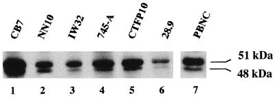
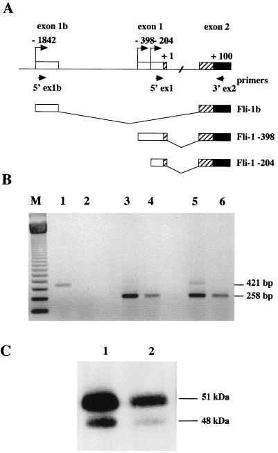
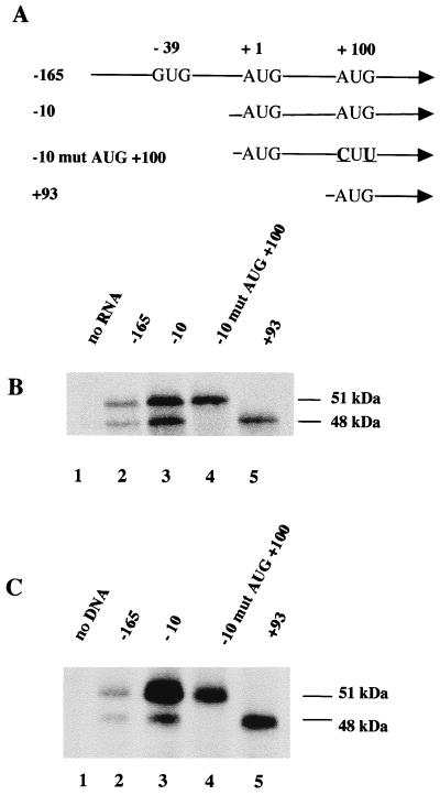
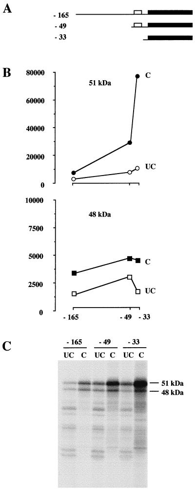
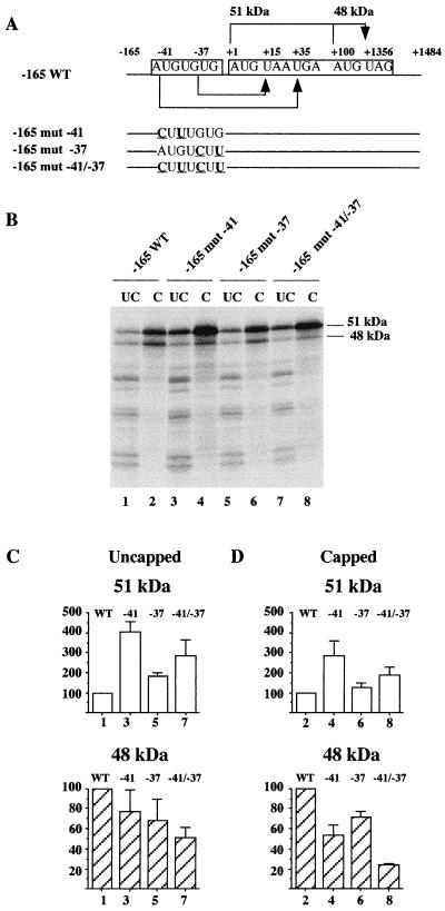
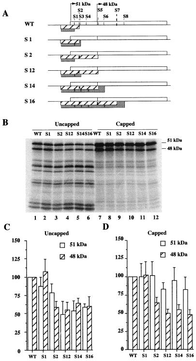
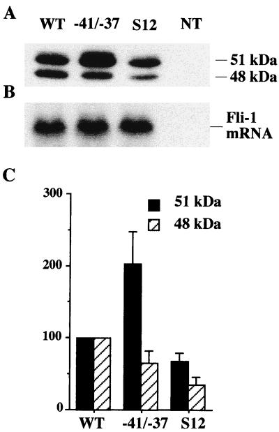
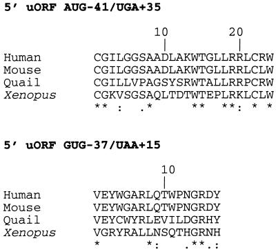
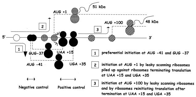
The proto-oncogene Fli-1 encodes a transcription factor of the ets family whose overexpression is associated with multiple virally induced leukemias in mouse, inhibits murine and avian erythroid cell differentiation, and induces drastic perturbations of early development in Xenopus. This study demonstrates the surprisingly sophisticated regulation of Fli-1 mRNA translation. We establish that two FLI-1 protein isoforms (of 51 and 48 kDa) detected by Western blotting in vivo are synthesized by alternative translation initiation through the use of two highly conserved in-frame initiation codons, AUG +1 and AUG +100. Furthermore, we show that the synthesis of these two FLI-1 isoforms is regulated by two short overlapping 5' upstream open reading frames (uORF) beginning at two highly conserved upstream initiation codons, AUG -41 and GUG -37, and terminating at two highly conserved stop codons, UGA +35 and UAA +15. The mutational analysis of these two 5' uORF revealed that each of them negatively regulates FLI-1 protein synthesis by precluding cap-dependent scanning to the 48- and 51-kDa AUG codons. Simultaneously, the translation termination of the two 5' uORF appears to enhance 48-kDa protein synthesis, by allowing downstream reinitiation at the 48-kDa AUG codon, and 51-kDa protein synthesis, by allowing scanning ribosomes to pile up and consequently allowing upstream initiation at the 51-kDa AUG codon. To our knowledge, this is the first example of a cellular mRNA displaying overlapping 5' uORF whose translation termination appears to be involved in the positive control of translation initiation at both downstream and upstream initiation codons.


Figures

FIG. 1
FIG. 1
Western blot analysis of FLI-1 proteins in total-cell lysates prepared from several Friend erythroleukemia cell lines (lanes 1 to 6) and from peripheral blood nucleated cells of a normal mouse (lane 7).
FIG. 2
FIG. 2
The mouse Fli-1b transcript is expressed in the leukemic pre-B-cell line 70Z/3 but not in the Friend erythroleukemia cell line 745-A. (A) Schematic representation of the 5′ genomic structure of the mouse Fli-1 gene and of the 5′ ends of known transcript isoforms. Coordinates of transcription initiation sites are given with respect to the last AUG located in exon 1 (AUG +1), usually described to initiate the large FLI-1 protein isoform. Open boxes represent 5′UTR sequences, and the solid box represents the common FLI-1 coding sequence of all Fli-1 transcript isoforms. The sequences which are alternatively coding (−398 and −204 transcripts) or noncoding (transcript Fli-1b) are indicated by hatched boxes. Small horizontal arrows indicate the position of the primers used in RT-PCR. (B) RT-PCR analysis of Fli-1 transcripts expressed in the pre-B-cell line 70Z/3 (lanes 1, 3, and 5) and in the erythroleukemia cell line 745-A (lanes 2, 4, and 6). All the RT-PCR amplifications were performed with equal amounts of total RNA and the common primer 3′ex2 with either primer 5′ex1b (lanes 1 and 2), primer 5′ex1 (lanes 3 and 4), or both (lanes 5 and 6). The lengths of specific Fli-1b cDNA (421 bp) or Fli-1 −398 and −204 cDNAs (258 bp) are given to the right. M, 123-bp ladder. (C) Control Western blot analysis of FLI-1 protein isoforms expressed in the pre-B-cell line 70Z/3 (lane 1) and the erythroleukemia cell line 745-A (lane 2).
FIG. 3
FIG. 3
Partial alignment of Fli-1 cDNA sequences from human (row 1), quail (row 2), mouse (row 3), and frog (row 4). Row C contains the consensus sequence. The coordinates of conserved translation initiation or termination codons (boxed) are given on top of the alignment. The phase of each conserved codon is indicated below the alignment.
FIG. 4
FIG. 4
Identification of the initiation codons involved in the synthesis of the 51- and 48-kDa FLI-1 proteins. (A) Schematic drawing of the 5′ ends of synthetic Fli-1 cDNA used to synthesize Fli-1 mRNA by in vitro transcription. Point mutations are underlined. (B) Equal amounts of each of these synthetic Fli-1 mRNA were used to program rabbit reticulocyte lysates in the presence of [35S]methionine. Translation products were then separated by SDS-PAGE, transferred to a nitrocellulose membrane, and revealed by autoradiography. (C) Western blot analysis of FLI-1 proteins synthesized in NIH 3T3 cells under transient expression of the transfected pCI Neo vector (Promega) carrying the indicated Fli-1 cDNA.
FIG. 5
FIG. 5
Differential regulation of the synthesis of the 51- and 48-kDa FLI-1 isoforms by the conserved region −49/−33. Capped and uncapped Fli-1 mRNAs harboring progressive deletions of the 5′UTR were synthesized by in vitro transcription. Equal amounts of each of these synthetic Fli-1 mRNA were used to program rabbit reticulocyte lysates in the presence of [35S]methionine. Translation products were separated by SDS-PAGE. Labeled translation products were then visualized by autoradiography, and radioactive signals corresponding to the 51- and 48-kDa proteins were quantified using a Molecular Imager. (A) Schematic representation of synthetic Fli-1 mRNA. The open boxes represent the −49/−33 conserved region in 5′UTR, and the solid boxes represent coding sequences. (B) Graphical representation of the results of the quantitation of the 51-kDa (top) or 48-kDa (bottom) isoforms synthesized with the capped (C) or uncapped (UC) versions of each Fli-1 mRNA. The y axis shows arbitrary units of radioactivity, and the x axis shows the type of mRNA used for translation. (C) Autoradiography of the gel.
FIG. 6
FIG. 6
Differential regulation of the synthesis of the 51- and 48-kDa FLI-1 isoforms by the conserved region −49/−33 is mediated by upstream codons, AUG −41 and GUG −37. Capped or uncapped versions of Fli-1 mRNA with or without mutations of AUG −41 or GUG −37 codons or with both mutations were synthesized by in vitro transcription. Equal amounts of each of these synthetic Fli-1 mRNAs were used to program rabbit reticulocyte lysates in the presence of [35S]methionine. Translation products were separated by SDS-PAGE. Labeled translation products were then visualized by autoradiography, and radioactive signals corresponding to the 51- and 48-kDa proteins were quantified using a Molecular Imager. (A) Schematic representation of synthetic Fli-1 mRNA. Point mutations are underlined. The two short 5′ uORFs beginning at AUG −41 or GUG −37 and ending, respectively, at UGA +35 and UAA +15 are indicated by arrows under the drawing of the wild-type mRNA. Similarly, the two in-frame FLI-1 ORF starting at AUG +1 or AUG +100 and ending at UAG +1356 are indicated by arrows above the drawing of the wild-type mRNA. (B) Autoradiogram of the gel. (C and D) Graphical representation of the results obtained with uncapped and capped versions of the same mRNA. Results are expressed as the percentage of 51-kDa (white bars) or 48-kDa (hatched bars) isoforms produced by the indicated mutated mRNA compared to the wild type (means and standard deviations of three different experiments).
FIG. 7
FIG. 7
Synthesis of both FLI-1 isoforms is positively regulated by the conserved UAA +15 and UGA +35 stop codons. The same experiment as in Fig. 6 was performed using the different mRNA shown in panel A, in which the overlap of the two 5′ uORFs initiated at either AUG −41 (hatched boxes) or GUG −37 (grey boxes) and the FLI-1 ORF (open boxes) was progressively increased by the mutations of the corresponding in-frame stop codons (crosses). These stop codons are numbered from 1 to 8 depending on their increasing distance from AUG +1. Stop codons S1 (UAA +15, changed to CAA), S3 (UGA +39, changed to CGA), S4 (UGA +57, changed to CGA) S6 (UGA +120, changed to CGA) and S8 (UGA +210) are located in the same frame as the GUG −37 codon, whereas stop codons S2 (UGA +35, changed to UCA), S5 (UGA +101, changed to UGC), and S7 (UGA +185) are located in the same frame as AUG −41 codon. (B) Autoradiogram of the gel showing the different proteins synthesized; the positions of the 51- and 48-kDa FLi-1 isoforms are indicated to the right. (C and D) Graphical representations of the quantitative analysis of the production of the 51-kDa and the 48-kDa isoforms from uncapped or capped versions of the same mRNA. Results are expressed as the percentage of the amounts of either 51- or 48-kDa isoforms produced by the indicated mRNA compared to the wild type (means and standard deviations of three different experiments).
FIG. 8
FIG. 8
Regulation of Fli-1 mRNA translation by conserved 5′ uORFs in vivo. NIH 3T3 cells were transfected by Fli-1 expression vectors carrying either the wild-type cDNA sequence (lane WT), mutations at both upstream AUG −41 and GUG −37 codons (lane −41/−37), or mutations at both stop codons UAA +15 and UGA +35 (lane S12). NT, nontransfected cells. (A) Western blot analysis of FLI-1 proteins produced in transfected cells. (B) Northern blot analysis of Fli-1 mRNA. (C) Quantitative analysis of the production of the 51- and 48-kDa proteins. Relative values are expressed as percentages of the amount of proteins produced by the wild-type sequence (means and standard deviations of four different transfections).
FIG. 9
FIG. 9
Alignment of the predicted amino acid sequences of the putative peptides encoded by the two conserved 5′ uORF in human, mouse, quail, and Xenopus mRNAs. Conserved amino acids are marked by asterisks.
FIG. 10
FIG. 10
Diagram illustrating the mechanisms involved in the regulation of Fli-1 mRNA translation by the conserved 5′ uORF. Step 1: Given the minimal twofold stimulation of the 51-kDa protein synthesis induced by the disruption of AUG −41 and GUG −37, we can estimate that at least half of the 40S subunits loaded at the 5′ end of Fli-1 mRNA initiate translation at upstream AUG −41 and GUG −37 codons. The remaining 40S subunits go on their 3′ progression by leaky scanning. Step 2: Translation initiation at AUG +1 is facilitated by the piling up of leaky-scanning 40S subunits against ribosomes terminating the translation of the two 5′ uORF at stop codons UAA +15 and UGA +35. Step 3: At least half of the translation initiated at AUG +100 is due to ribosomes which reinitiate after translation termination at stop codons UAA +15 and UGA +35, while the remaining translation is due to scanning 40S subunits having bypassed all upstream initiation codons.

References

    1. Baily R A, Bosselut R, Zucman J, Cormier F, Delattre O, Roussel M, Thomas G, Ghysdael J. DNA-binding and transcriptional activation properties of the EWS-FLI-1 fusion protein resulting from the t(11;22) translocation in Ewing sarcoma. Mol Cell Biol. 1994;14:3230–3241. - PMC - PubMed
    1. Barbeau B, Bergeron D, Beaulieu M, Nadjem Z, Rassart E. Characterization of the human and mouse Fli-1 promoter regions. Biochim Biophys Acta. 1996;1307:220–232. - PubMed
    1. Ben-David Y, Giddens E R, Bernstein A. Identification and mapping of a common proviral integration site Fli1 in erythroleukemia cells induced by Friend murine leukemia virus. Proc Natl Acad Sci USA. 1990;87:1332–1336. - PMC - PubMed
    1. Ben-David Y, Bernstein A. Friend virus induced erythroleukemia and the multistage of cancer. Cell. 1991;66:831–834. - PubMed
    1. Bergeron D, Poliquin L, Houde J, Barbeau B, Rassart E. Analysis of proviruses integrated in Fli-1 and Evi-1 regions in Cas-Br-E MuLV-induced non-T-, non-B-cell leukemias. Virology. 1992;191:661–669. - PubMed

Publication types

MeSH terms

LinkOut - more resources