. 2000 May;20(9):2984-95.
doi: 10.1128/MCB.20.9.2984-2995.2000.

A novel cold-sensitive allele of the rate-limiting enzyme of fatty acid synthesis, acetyl coenzyme A carboxylase, affects the morphology of the yeast vacuole through acylation of Vac8p

Affiliations

A novel cold-sensitive allele of the rate-limiting enzyme of fatty acid synthesis, acetyl coenzyme A carboxylase, affects the morphology of the yeast vacuole through acylation of Vac8p

R Schneiter et al. Mol Cell Biol. 2000 May.

Abstract

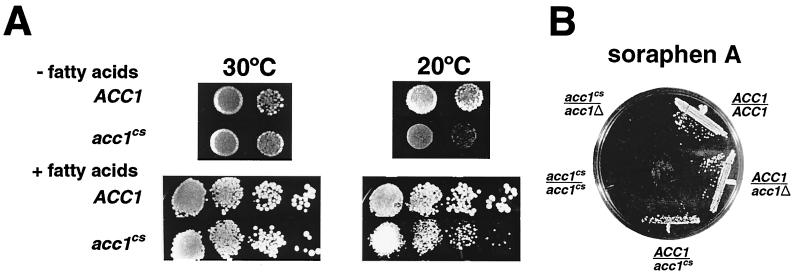
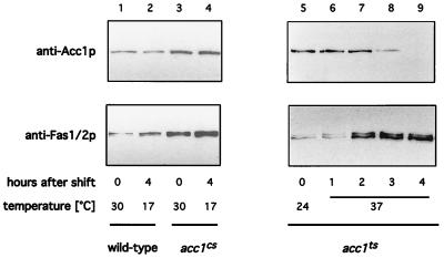
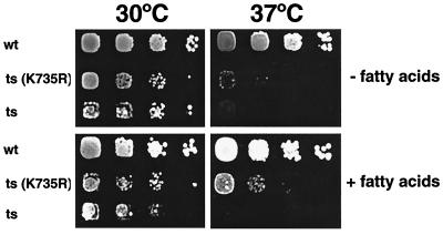
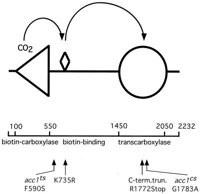
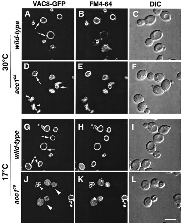
The yeast vacuole functions both as a degradative organelle and as a storage depot for small molecules and ions. Vacuoles are dynamic reticular structures that appear to alternately fuse and fragment as a function of growth stage and environment. Vac8p, an armadillo repeat-containing protein, has previously been shown to function both in vacuolar inheritance and in protein targeting from the cytoplasm to the vacuole. Both myristoylation and palmitoylation of Vac8p are required for its efficient localization to the vacuolar membrane (Y.-X. Wang, N. L. Catlett, and L. S. Weisman, J. Cell Biol. 140:1063-1074, 1998). We report that mutants with conditional defects in the rate-limiting enzyme of fatty acid synthesis, acetyl coenzyme A carboxylase (ACC1), display unusually multilobed vacuoles, similar to those observed in vac8 mutant cells. This vacuolar phenotype of acc1 mutant cells was shown biochemically to be accompanied by a reduced acylation of Vac8p which was alleviated by fatty acid supplementation. Consistent with the proposed defect of acc1 mutant cells in acylation of Vac8p, vacuolar membrane localization of Vac8p was impaired upon shifting acc1 mutant cells to nonpermissive condition. The function of Vac8p in protein targeting, on the other hand, was not affected under these conditions. These observations link fatty acid synthesis and availability to direct morphological alterations of an organellar membrane.


Figures

FIG. 1
FIG. 1
Effects of fatty acids and of soraphen A on growth of the acc1cs mutant. (A) Haploid yeast colonies of the indicated genotype were resuspended in water, and aliquots of dilutions were plated on medium with and without fatty acid supplementation; 10 μl containing ca. 104, 103, 102, or 101 cells was spotted onto plates, which were incubated for 3 days at 30°C or for 7 days at 20°C. (B) The soraphen A-sensitive phenotype of acc1cs is semidominant. Diploid strains of indicated genotypes were streaked onto YEPD medium containing 0.25 μg of soraphen A per ml, and the plate was incubated for 2 days at 30°C.
FIG. 2
FIG. 2
Steady-state levels of Acc1p in conditional acc1 mutants, determined by Western blotting to detect Acc1p and Fas1/2p expression in wild-type (W303; lane 1 and 2) acc1cs (479-2A; lane 3 and 4), and acc1ts (YRXS12; lanes 5 to 9) strains at the indicated times at permissive and nonpermissive conditions.
FIG. 3
FIG. 3
A biotinylation-deficient allele of ACC1, acc1K735R, partially rescues the acc1ts allele. Wild-type (wt; W303), the acc1ts mutant strain (ts; YRXS12), and the acc1ts mutant harboring a plasmid encoding the biotinylation deficient allele of ACC1 [ts(K735R); pRXS89] were serially diluted 10-fold and spotted onto YEPD and YEPD plates supplemented with fatty acids. Plates were incubated for 3 days at 30 or 37°C.
FIG. 4
FIG. 4
Map (drawn to scale) summarizing the positions of mutations in different acc1 alleles. The biotin prosthetic group is represented by a diamond-shaped symbol, the biotin-carboxylase domain is represented by a triangle (positions 100-550), and the transcarboxylase domain is denoted by a circle (positions 1450 to 2050). Lesions associated with different alleles are indicated.
FIG. 5
FIG. 5
Acc1C-termp stabilizes the thermolabile enzymatic complex formed by Acc1tsp. For Western blot analysis of acc1ts (YRXS12) transformed with an empty vector or with pCG002::Tn10-LUK#21 encoding the C-terminally truncated version of Acc1p, cells were grown at permissive conditions in synthetic medium lacking leucine and shifted to nonpermissive conditions for 8 h. Protein extracts were prepared and separated by SDS-PAGE, and blots were probed with peroxidase-conjugated ExtrAvidin to detect biotinylated Acc1p and the two isoforms of pyruvate carboxylase (Pyc1/2p).
FIG. 6
FIG. 6
Morphological analysis of acc1cs mutant cells. Transmission electron micrographs show wild-type (wt; A) and acc1cs mutant cells (B to D) shifted to nonpermissive conditions for 4 h in the absence (B and D) or presence (C) of supplemented fatty acids (fa). Fragmented vacuoles are indicated by arrows in panel B. Electron-dense granular structures reminiscent of Acc1p filaments are indicated by white stars in panel D. Bars: A to C, 1 μm; D, 0.1 μm.
FIG. 7
FIG. 7
Vacuolar morphology in wild-type, acc1cs, and acc1ts cells stained with FM4-64 Wild-type, acc1cs, and acc1ts cells were cultivated in YEPD or YEPD supplemented with fatty acids to early logarithmic growth phase at 30°C. Cells were then incubated with 30 μM FM4-64 for 30 min, followed by a chase for 1 h in YEPD or YEPD supplemented with fatty acids at 30°C. They were then incubated at the permissive temperature (30°C) or shifted to nonpermissive conditions (17°C for 3 h or 37°C for 30 min) and examined by confocal microscopy. Multilobed vacuoles are indicated by arrowheads in panels E, M, and Q. Dead cells in panels V and X are indicated by arrows. DIC (differential interference contrast) pictures of the visual fields are shown to the right of the fluorescence images. Bar, 10 μm.
FIG. 8
FIG. 8
Membrane association and acylation of Vac8p, but not processing of API, is affected in acc1cs mutant cells. (A) Membranes isolated from exponentially growing wild-type, vac8-3 (expressing a nonpalmitoylatable allele of Vac8p) (52), and acc1cs cells were subfractionated into P100 and S100 fractions, and membrane association of Vac8p was determined by immunoblot analysis with an anti-Vac8p serum. (B) Whole cell extracts of wild-type (wt) and cs mutant cells shifted to nonpermissive conditions for 4 h in the presence (+fa) or absence of exogenously added fatty acids were separated by SDS-PAGE, and the relative mobility of Vac8p was assessed by immunoblot analysis with an anti-Vac8p serum. (C) API processing in wild-type (wt), vac8Δ, vac8-3, and acc1cs mutant cells (cs) incubated at either permissive (30°C) or nonpermissive (17°C) conditions for 4 h was determined by immunoblot analysis with an anti-API serum. The positions of the precursor (p) and mature (m) forms of API are indicated.
FIG. 9
FIG. 9
Subcellular distribution of Vac8p fused to GFP in wild-type and acc1cs mutant cells. Wild-type and acc1cs mutant cells expressing a functional Vac8p-GFP fusion protein were grown to early logarithmic growth phase at 30°C. Cells were then incubated with 30 μM FM4-64 for 30 min, followed by a chase for 1 h in YEPD. The cultures were split and incubated at either permissive (30°C) or nonpermissive (17°C) conditions for 4 h. Vacuolar morphology and the subcellular distribution of Vac8p-GFP was then analyzed by confocal microscopy. The polarized distribution of Vac8p-GFP on the relaxed vacuolar membrane of wild-type cells is indicated by arrows in panels A, D, and G. Small arrowheads in panels D and J point to a polarized distribution of Vac8p-GFP on multilobed vacuolar structures. Aberrant distribution of Vac8p-GFP in acc1cs mutant cells at nonpermissive conditions is indicated by larger arrowheads in panel J. DIC (differential interference contrast) pictures of the visual fields are shown to the right of the fluorescence images. Bar, 5 μm.

References

    1. Aguilera A, Klein H L. Genetic and molecular analysis of recombination events in Saccharomyces cerevisiae occurring in the presence of the hyper-recombination mutation hpr1. Genetics. 1989;122:503–517. - PMC - PubMed
    1. Aguilera A, Klein H L. Genetic control of intrachromosomal recombination in Saccharomyces cerevisiae. I. Isolation and genetic characterization of hyper-recombination mutations. Genetics. 1988;119:779–790. - PMC - PubMed
    1. Aguilera A, Klein H L. HPR1, a novel yeast gene that prevents intrachromosomal excision recombination, shows carboxy-terminal homology to the Saccharomyces cerevisiae TOP1 gene. Mol Cell Biol. 1990;10:1439–1451. - PMC - PubMed
    1. Al-Feel W, Chirala S S, Wakil S J. Cloning of the yeast FAS3 gene and primary structure of yeast acetyl-CoA carboxylase. Proc Natl Acad Sci USA. 1992;89:4534–4538. - PMC - PubMed
    1. Brown D A, London E. Functions of lipid rafts in biological membranes. Annu Rev Cell Dev Biol. 1998;14:111–136. - PubMed

Publication types

MeSH terms