. 2000 May;20(9):2996-3003.
doi: 10.1128/MCB.20.9.2996-3003.2000.

Ku recruits the XRCC4-ligase IV complex to DNA ends

Affiliations

Ku recruits the XRCC4-ligase IV complex to DNA ends

S A Nick McElhinny et al. Mol Cell Biol. 2000 May.

Abstract

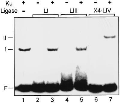
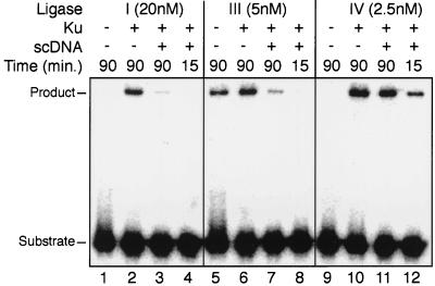
Genetic experiments have determined that Ku, XRCC4, and ligase IV are required for repair of double-strand breaks by the end-joining pathway. The last two factors form a tight complex in cells. However, ligase IV is only one of three known mammalian ligases and is intrinsically the least active in intermolecular ligation; thus, the biochemical basis for requiring this ligase has been unclear. We demonstrate here a direct physical interaction between the XRCC4-ligase IV complex and Ku. This interaction is stimulated once Ku binds to DNA ends. Since XRCC4-ligase IV alone has very low DNA binding activity, Ku is required for effective recruitment of this ligase to DNA ends. We further show that this recruitment is critical for efficient end-joining activity in vitro. Preformation of a complex containing Ku and XRCC4-ligase IV increases the initial ligation rate 20-fold, indicating that recruitment of the ligase is an important limiting step in intermolecular ligation. Recruitment by Ku also allows XRCC4-ligase IV to use Ku's high affinity for DNA ends to rapidly locate and ligate ends in an excess of unbroken DNA, a necessity for end joining in cells. These properties are conferred only on ligase IV, because Ku does not similarly interact with the other mammalian ligases. We have therefore defined cell-free conditions that reflect the genetic requirement for ligase IV in cellular end joining and consequently can explain in molecular terms why this factor is required.


Figures

FIG. 1
FIG. 1
Purification of X4-LIV. (A) SDS-PAGE gel of protein fractions stained with Coomassie blue. Lane 1, total soluble extract (xt); lane 2, peak fractions from Ni column; lane 3, peak fractions from Mono Q column; lane 4, molecular weight marker (M). X4, XRCC4, LIV, ligase IV. (B) Gel filtration of purified X4-LIV. A280, UV absorbance at 280 nm; Ve, volume eluted after injection of sample. (C) Elution plot of X4-LIV relative to mass standards (in kilodaltons). Vo, excluded volume for this column. The Ve/Vo ratios for five molecular mass standards are marked by diamonds. The determined Ve/Vo ratio for X4-LIV is noted relative to a line derived from regression of the five standards.
FIG. 2
FIG. 2
EMSA of Ku and X4-LIV. 5′ 32P-labeled 60-bp DNA duplex (50 nM) was present in all reactions. F, free DNA probe. Species I and II are DNA-protein complexes. (A) Formation of DNA-protein complexes with Ku and X4-LIV. The concentration of Ku, when present (+), was 5 nM. X4-LIV was added to a concentration of 50 (lanes 2 and 3), 25 (lane 4), 12.5 (lane 5), 5 (lane 6), and 2.5 nM (lane 7). (B) Characterization of DNA-protein complexes with antibodies. Ku (5 nM) was present in all reactions. The concentration of X4-LIV when present was 25 nM. αX4, polyclonal antiserum to XRCC4; NRS, normal rabbit serum; αKu, monoclonal antibody to Ku; αSV, monoclonal antibody to SV40 T antigen.
FIG. 3
FIG. 3
Detection of protein complexes by immunoprecipitation. All reactions contained 25 nM 32P-labeled X4-LIV. When present (+), Ku was at 5 nM. DNA species included in the reactions were e, a 60-bp duplex at 25 nM (0.5 μg), and sc, 0.5 μg of a supercoiled plasmid. Ab, antibody. Protein A-Sepharose and a monoclonal antibody to Ku (αKu) or an isotype-matched control antibody to SV40 T antigen (αSV; lane 1) were used to precipitate DNA-protein complexes. (A) Coomassie blue-stained gel of precipitated complexes. Ku80, 83-kDa subunit of Ku; Ku70, 70-kDa subunit of Ku. BSA was present due to its inclusion in the wash buffer. (B) Phosphorimage of dried gel. L-IV, ligase IV.
FIG. 4
FIG. 4
Immunoprecipitation of Ku from HeLa cell extracts. A 20-ng aliquot of the appropriate recombinant antigen (rAg; recombinant 70-kDa subunit of Ku or recombinant ligase IV), a 20-μg aliquot of the input extract (xt), and material recovered from immunoprecipitations (IPs) were electrophoresed and blotted onto a nitrocellulose membrane. αKu, immunoprecipitation with a monoclonal antibody to Ku; αSV, immunoprecipitation with an isotype-matched monoclonal antibody to SV40 T antigen; +EtBr, immunoprecipitation supplemented with 50 μg of ethidium bromide/ml. Recombinant antigens migrate slightly slower than the native antigens from HeLa cells due to the presence of C-terminal hexahistidine tags. Immunoblotting with the appropriate polyclonal antisera was used to detect Ku70 (A) and ligase IV (B).
FIG. 5
FIG. 5
Functional test for formation of Ku-X4-LIV complexes. (A) Competition assay design. Substrate L is a pair of dsDNA molecules. One dsDNA is 5′ 32P labeled on a 60-nucleotide (nt) strand, which can be ligated only to the 36-nt strand of a second dsDNA due to complementary 2-bp 3′ overhangs. Substrate S is another pair of dsDNA molecules. One dsDNA is 5′ 32P labeled on a 58-nt strand that can be ligated only to a 36-nt strand of a second dsDNA due to a different pair of complementary 2-bp 3′ overhangs. Asterisks show the locations of a 5′ 32P label. Step 1, substrates are preincubated on ice for 20 min, with or without Ku and/or X4-LIV, varied for each of the three reactions as noted. Step 2, preincubations are mixed together. In reaction I only, Ku is added after mixing. All three reactions are now equivalent and contain a final concentration of 5 nM for each of the four DNA molecules, 2.5 nM X4-LIV, and 10 nM Ku. Step 3, ligation is initiated by addition of 5 mM Mg2+ and 0.1 mM ATP and incubation at 37°C. Step 4, aliquots of each reaction are taken at successive time points and analyzed by denaturing gel electrophoresis. (B) Results of competition assay. Reactions I, II, and III were assembled as described above. Time is noted in minutes after the start of the reaction. The positions of the labeled strands of the substrates and products for both L and S pairs are marked. (C) Effect of preincubation on reaction kinetics. Reaction mixtures were preincubated with substrate L (described above; 5 nM each duplex) and (i) 5 nM Ku, and 2.5 nM X4-LIV, (ii) 2.5 nM X4-LIV only, (iii) 5 nM Ku only, or (iv) no protein (No Pre.) for 20 min on ice. Ku and/or X4-LIV was then added so that all four reaction mixtures were equivalent, and the reactions were initiated by the addition of Mg2+. Aliquots of each reaction mixture were taken at the indicated times (in minutes), and the ligation product was quantified as a percentage of the total starting substrate.
FIG. 6
FIG. 6
Specificity of complex formation. A 5′ 32P-labeled 60-bp DNA duplex was present in all reactions at a concentration of 50 nM. F, free DNA probe. The mobilities of DNA-protein complexes are noted for species I and II. When present (+), Ku was added to a concentration of 5 nM. LI, 50 nM ligase I; LIII, 50 nM ligase III; X4-LIV, 25 nM XRCC4 plus ligase IV.
FIG. 7
FIG. 7
Effect of unbroken competitor DNA on activities of different ligases. Substrate L (Fig. 5A) was present in all reactions at 5 nM for each of the two duplex DNAs. Ku, when present (+), was added to a concentration of 10 nM and preincubated with the ligation substrate for 15 min at 25°C. Two micrograms of supercoiled DNA was added when noted, and ligation was initiated by the subsequent addition of 20 nM ligase I (lanes 1 to 4), 5 nM ligase III (lanes 5 to 8), or 2.5 nM X4-LIV (lanes 9 to 12). Reaction mixtures were incubated for the indicated times (in minutes) at 37°C, and product formation was assessed by denaturing gel electrophoresis.

References

    1. Baumann P, West S C. DNA end-joining catalyzed by human cell-free extracts. Proc Natl Acad Sci USA. 1998;95:14066–14070. - PMC - PubMed
    1. Bogue M A, Jhappan C, Roth D B. Analysis of variable (diversity) joining recombination in DNA-dependent protein kinase (DNA-PK)-deficient mice reveals DNA-PK-independent pathways for both signal and coding joint formation. Proc Natl Acad Sci USA. 1998;95:15559–15564. - PMC - PubMed
    1. Bogue M A, Wang C, Zhu C, Roth D B. V(D)J recombination in Ku86-deficient mice: distinct effects on coding, signal, and hybrid joint formation. Immunity. 1997;7:37–47. - PubMed
    1. Bryans M, Valenzano M C, Stamato T D. Absence of DNA ligase IV protein in XR-1 cells: evidence for stabilization by XRCC4. Mutat Res. 1999;433:53–58. - PubMed
    1. Cary R B, Peterson S R, Wang J, Bear D G, Bradbury E M, Chen D J. DNA looping by Ku and the DNA-dependent protein kinase. Proc Natl Acad Sci USA. 1997;94:4267–4272. - PMC - PubMed

Publication types