. 2000 May;20(9):3015-26.
doi: 10.1128/MCB.20.9.3015-3026.2000.

TAK1 participates in c-Jun N-terminal kinase signaling during Drosophila development

Affiliations

TAK1 participates in c-Jun N-terminal kinase signaling during Drosophila development

Y Takatsu et al. Mol Cell Biol. 2000 May.

Abstract

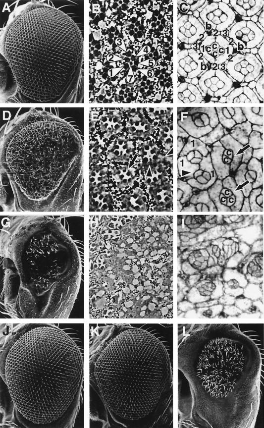
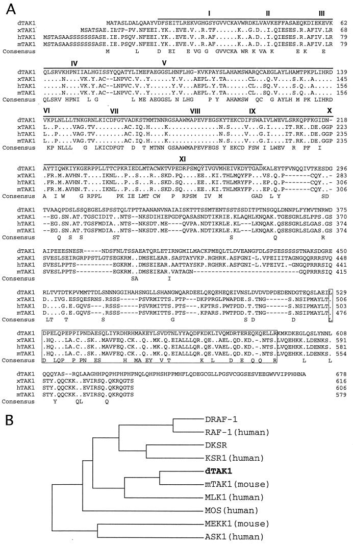
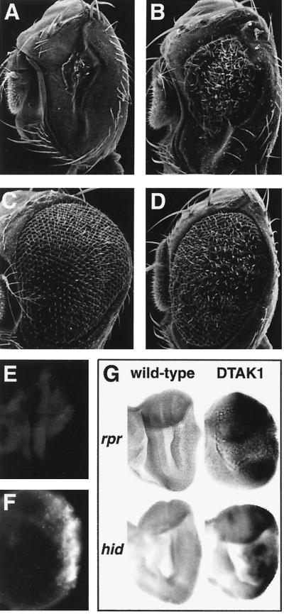
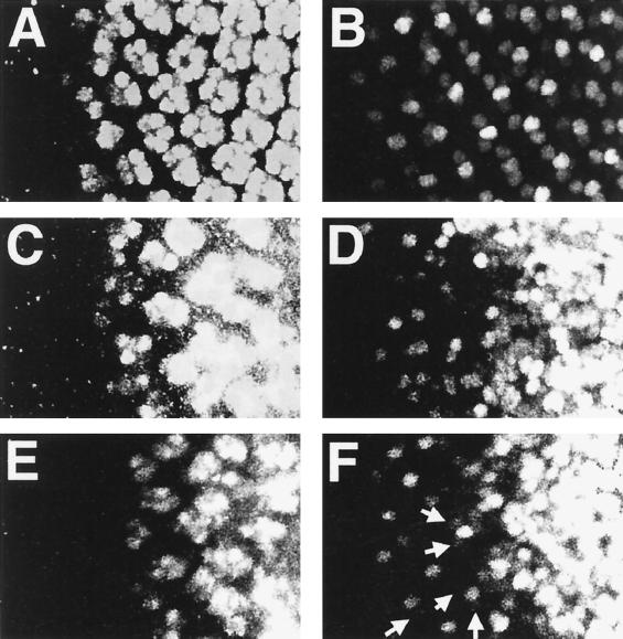
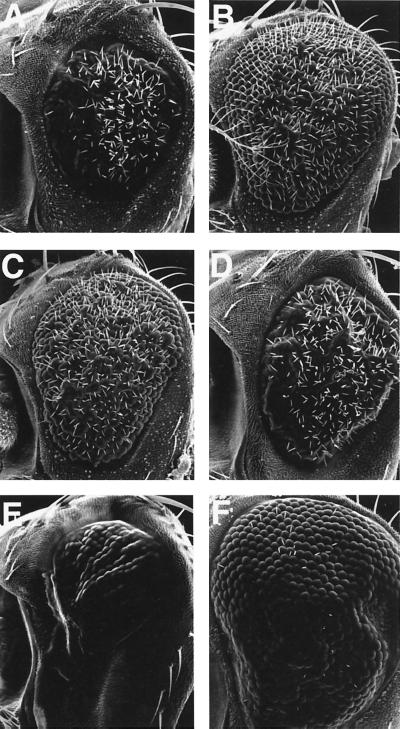
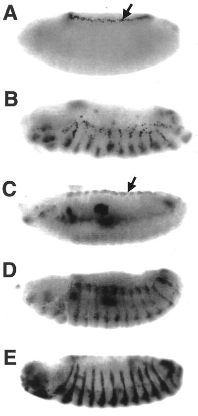
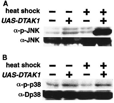
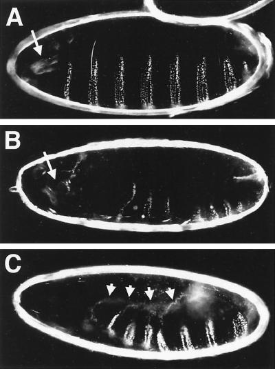
Transforming growth factor beta (TGF-beta)-activated kinase 1 (TAK1) is a member of the MAPKKK superfamily and has been characterized as a component of the TGF-beta/bone morphogenetic protein signaling pathway. TAK1 function has been extensively studied in cultured cells, but its in vivo function is not fully understood. In this study, we isolated a Drosophila homolog of TAK1 (dTAK1) which contains an extensively conserved NH(2)-terminal kinase domain and a partially conserved COOH-terminal domain. To learn about possible endogenous roles of TAK1 during animal development, we generated transgenic flies which express dTAK1 or the mouse TAK1 (mTAK1) gene in the fly visual system. Ectopic activation of TAK1 signaling leads to a small eye phenotype, and genetic analysis reveals that this phenotype is a result of ectopically induced apoptosis. Genetic and biochemical analyses also indicate that the c-Jun amino-terminal kinase (JNK) signaling pathway is specifically activated by TAK1 signaling. Expression of a dominant negative form of dTAK during embryonic development resulted in various embryonic cuticle defects including dorsal open phenotypes. Our results strongly suggest that in Drosophila melanogaster, TAK1 functions as a MAPKKK in the JNK signaling pathway and participates in such diverse roles as control of cell shape and regulation of apoptosis.


Figures

FIG. 1
FIG. 1
Phenotypes induced by ectopic TAK1 signaling. Scanning electron micrographs of the compound eye (A, D, G, J, K, and L), tangential histological sections (B, E, and H), and pupal eyes (at 40 h after puparium formation) stained with cobalt sulfide (C, F, and I) are shown. (A to C) Canton-S. (A) The wild-type eye is composed of a regular array of about 800 ommatidia. (B) Each ommatidium contains six outer photoreceptor cells and an inner photoreceptor cell, R7 (R1 to R7 are indicated with numbers). (C) Cobalt sulfide staining shows the apical profile of cells in the epithelium. Each ommatidium contains four cone cells (indicated with “c”) and two primary pigment cells (“1”), surrounded by six secondary pigment cells (“2”), three tertiary pigment cells (“3”), and three interommatidial bristles (“b”). (D to F) pGMR-mTAK overexpression. (E) Most of the ommatidia contain only five to six photoreceptors (arrow), and visual pigments are also disrupted at many positions (arrowheads). (F) In the pupal eye, most of the ommatidia contain only two or three cone cells (arrows) and occasionally are also missing primary pigment cells (arrowhead). Numerous numbers of interommatidial cells are also missing in this mutant. (G to I) pGMR-mTAK1ΔN overexpression. (G) Expression of mTAK1ΔN in the developing eye results in a severe decrease in size of the compound eyes. (H and I) Ommatidial structures are totally disrupted and hard to discriminate in the adult head section and pupal eye disc. (J) pGMR-hTAB1. Expression of hTAB1 alone in the eye has no effect on eye development. (K) Weak pGMR-mTAK1 overexpression phenotype. (L) Coexpression of hTAB1 and mTAK1 (weak line as shown in panel K) causes a phenotype as severe as pGMR-mTAK1ΔN overexpression, as shown in panel G. All pictures are shown with anterior to the left and dorsal up.
FIG. 2
FIG. 2
Primary structure of dTAK1. (A) The dTAK1 primary sequence is compared with that of xTAK1, one of the three alternative splicing forms of hTAK1 (hTAK1b), and mTAK1. The protein sequences are presented in single-letter code. Gaps (−) were introduced to optimize the alignment. Identical residues are indicated with periods. The protein kinase domain sequence is shown by overline, and sequences corresponding to conserved kinase subdomains I to XI (27) are indicated by roman numerals. The 65-residue stretch of amino acids in the COOH-terminal domain that is conserved between TAK1s is boxed. (B) Relationship between catalytic domains of members of the vertebrate and Drosophila MAPKKK group, presented as a dendrogram created using the Gene Works program (version 2.0; IntelliGenetics). The figure presents the analysis of the human MAPKKKs RAF-1, KSR1, MLK1, MOS, and ASK1 (3, 14, 34, 63, 68), the mouse MAPKKKs TAK1 and MEKK1 (40, 71), and the Drosophila MAPKKKs DRAF-1, DKSR (48, 63), and dTAK1.
FIG. 3
FIG. 3
Ectopic TAK1 signaling induces apoptosis in the developing eye. (A) GMR-GAL4/UAS-dTAK1(strong); (B) GMR-GAL4/UAS-dTAK1(weak); (C) GMR-GAL4/UAS-dTAK1(weak); pGMR-p35; (D) GMR-GAL4/UAS-dTAK1(weak); Df(3L)H99/+. The deficiency of Df(3L)H99 uncovers three proapoptotic genes, rpr, hid, and grim. The reduced eye phenotype of GMR-GAL4/UAS-dTAK1(weak) (B) is rescued either by coexpression of p35 (C) or by a heterozygous mutant background which removes the proapoptotic genes (D). (E and F) Acridine orange staining of eye discs at late third-instar larval stage of Canton-S (E) and GMR-GAL4/UAS-dTAK1 (F) flies. Acridine orange-positive cells are rare in the wild-type eye disc (E) but abundant in dTAK1 overexpression eye discs predominantly posterior to the morphogenetic furrow (F). (G) Eye discs from wild-type flies (left two discs) or GMR-GAL4/UAS-dTAK1 flies (right) were labeled with an rpr antisense riboprobe (top) or a hid antisense riboprobe (bottom). rpr and hid expression is not evident in the wild-type eye disc. dTAK1 expression induces rpr and hid most significantly in regions posterior to the morphogenetic furrow.
FIG. 4
FIG. 4
Effect of dTAK1 expression in photoreceptor cell induction. Late third-instar eye discs were doubly labeled with anti-Elav antibody (A, C, E) and for rhomboid lacZ (X81) expression. Anti-Elav antibody stains all of the photoreceptor cells (R1 to R8). X81 expresses lacZ strongly in R8 and relatively weakly in R2 and R5 (19). (A and B) Wild type; (C and D) GMR-GAL4/UAS-dTAK1; (E and F) GMR-GAL4/UAS-dTAK1; pGMR-p35. R8 induction occurs normal in GMR-GAL4/UAS-dTAK1 (D) and GMR-GAL4/UAS-dTAK1; pGMR-p35 (F) eye discs. R2 and R5 are occasionally induced in the GMR-GAL4/UAS-dTAK1; pGMR-p35 disc (indicated with arrows in panel F). Photoreceptor cell markers are disordered and diffuse in more posterior regions (C to F). This result indicates that dTAK1 overexpression altered or delayed specification of the photoreceptor cells, particularly for R3, R4, R1, R6, and R7. All images are presented with anterior to the left.
FIG. 5
FIG. 5
Genetic interaction of pGMR-mTAK1ΔN. (A to F) Scanning electron micrographs of the compound eye. (A) GMR-GAL4/UAS-dTAK1(weak); (B) hep1/Y; GMR-GAL4/UAS-dTAK1(weak); (C) GMR-GAL4/UAS-dTAK1(weak); bsk2/+; (D) Dsor1LH110/Y; GMR-GAL4/UAS-dTAK1(weak) (E) GMR-GAL4/+; UAS-hep/+; (F) GMR-GAL4/+; UAS-hep/pGMR-p35. Reduced eye phenotype of GMR-GAL4/UAS-dTAK1 (A) is suppressed by one copy reduction of the hep or bsk gene (B and C). In contrast to this, a mutant which is involved in MAPK/ERK cascade, Dsor1, does not show any genetic interaction to this phenotype (D). Ectopic expression of hep also results in the small eye phenotype (E). This phenotype is suppressed by the presence of p35 (F), indicating that ectopic activation of the JNK signal induced apoptosis in the developing eye. GMR-GAL4/UAS-hep flies lost most of the interommatidial bristles (E). Bristle phenotype is not rescued by p35 expression (F), suggesting that apoptosis is not a direct cause of this phenotype. Anterior is to the left, and dorsal is up.
FIG. 6
FIG. 6
Ectopic induction of puc and dpp by dTAK1 demonstrated by X-Gal staining (A and B) and in situ hybridization for dpp antisense probe (C and D) of stage 14 embryos. (A) puc-lacZ/+; (B) en-GAL4/UAS-dTAK1; puc-lacZ/+; (C) wild type; (D) en-GALY/UAS-dTAK1. puc and dpp expression in the leading-edge cells is indicated by arrows (A and C, respectively). Ectopic expression of dTAK1, controlled by en-GAL4, ectopically induces both puc and dpp in the embryonic ectoderm with a striped pattern (B and D). (E) en-GAL4/UAS-lacZ. en-GAL4 expression pattern is shown. Anterior is to the left, and dorsal is up.
FIG. 7
FIG. 7
In vivo phosphorylation of Bsk and D-p38 by ectopic dTAK1 expression. Third-instar larva carrying only hs-GAL4 or both UAS-dTAK1 and hs-GAL4 were collected with or without heat shock treatment. Extracts prepared from these larva were immunoblotted with either anti-p-JNK or anti-JNK1 antibody (A) and with anti-p-D-p38 or anti-p38 (B). Bsk phosphorylation is increased dramatically only in UAS-dTAK1-carrying animals upon heat shock. However, phosphorylation of D-p38 is not induced by the dTAK1 overexpression.
FIG. 8
FIG. 8
Lateral view of the cuticle phenotypes of dominant negative dTAK1-expressing embryos of the following genotypes: (A) +/pannierMD237GAL4, as a wild-type control; (B and C) UAS-dTAK1-K46R/+; UAS-dTAK1-K46R/pannierMD237GAL4. (A) Wild-type cuticle illustrating the regular spacing of the denticle belt on the ventral side and complete closure of the epidermis on the dorsal side. (B and C) Expression of dTAK1-K46R (two copies of transgene) during embryonic development by means of pnr-GAL4 (32) causes various defects. Defects in the anterior structure, typically loss of the mouth hooks (normal position of the mouth hooks is indicated with arrows in panels A and B), are seen in 37% of embryos (n = 591). Embryos of this type are frequently exhibit a small whole in the anterior and dorsal side of the cuticle (B). In the most extreme cases, the embryo is completely open dorsally (6%, n = 591) (C). Arrows indicate the edge of the dorsal hole of the cuticle.
FIG. 9
FIG. 9
Phenotypes induced by the expression of dominant negative TAK1. Pupal eyes of the following genotypes at 40 h after puparium formation were stained with cobalt sulfide. (A) pGMR-mTAK1-K63W (one copy). Expression of mTAK1-K63W, a dominant negative form of mTAK1, at a lower level results in defective positioning of the interommatidial bristle (indicated with arrows, compared to the wild-type shown in Fig. 1C). (B) pGMR-mTAK1-K63W (two copies). A higher level of mTAK1-K63W expression totally disrupts the ommatidial array. The cell shapes of secondary and tertiary pigment cells are irregular, and it is hard to discriminate these two cell types by morphology. (C) GMR-GAL4/UAS-dTAK1-K46R; UAS-dTAK1-K46R/+. A similar phenotype is observed in a fly expressing dTAK1-K46R (two copies), a kinase-inactive form of dTAK1. (D) hepr75/Y. The hep mutant disc also shows bristle mislocation (indicated with arrows) and abnormal pigment cell shapes (arrowheads). All images show the phenotype of the center region of the pupal eye discs (even in the wild-type discs, bristle mislocation is occasionally observed in the anterior edge region). Anterior is to the left.

References

    1. Adachi-Yamada T, Nakamura M, Irie K, Tomoyasu Y, Sano Y, Mori E, Goto S, Ueno N, Nishida Y, Matsumoto K. p38 mitogen-activated protein kinase can be involved in transforming growth factor β superfamily signal transduction in Drosophila wing morphogenesis. Mol Cell Biol. 1999;19:2322–2329. - PMC - PubMed
    1. Ashburner M. Drosophila: a laboratory manual. New York, N.Y: Cold Spring Harbor Laboratory Press; 1989.
    1. Bonner T I, Oppermann H, Seeburg P, Kerby S B, Gunnell M A, Young A C, Rapp U R. The complete coding sequence of the human raf oncogene and the corresponding structure of the c-raf-1 gene. Nucleic Acids Res. 1986;14:1009–1015. - PMC - PubMed
    1. Boutros M, Paricio N, Strutt D I, Mlodzik M. Dishevelled activates JNK and discriminates between JNK pathways in planar polarity and wingless signaling. Cell. 1998;94:109–118. - PubMed
    1. Bowtell D D, Kimmel B E, Simon M A, Rubin G M. Regulation of the complex pattern of sevenless expression in the developing Drosophila eye. Proc Natl Acad Sci USA. 1989;86:6245–6249. - PMC - PubMed

Publication types

MeSH terms

Substances

Associated data