. 2000 May;20(9):3137-46.
doi: 10.1128/MCB.20.9.3137-3146.2000.

The human SWI-SNF complex protein p270 is an ARID family member with non-sequence-specific DNA binding activity

Affiliations

The human SWI-SNF complex protein p270 is an ARID family member with non-sequence-specific DNA binding activity

P B Dallas et al. Mol Cell Biol. 2000 May.

Abstract

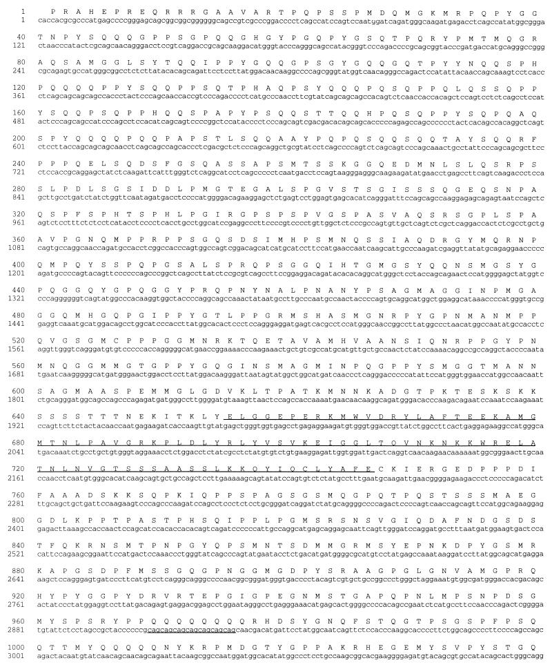
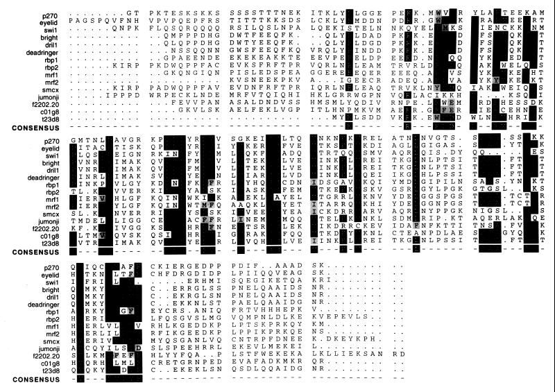
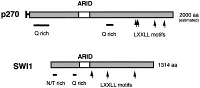
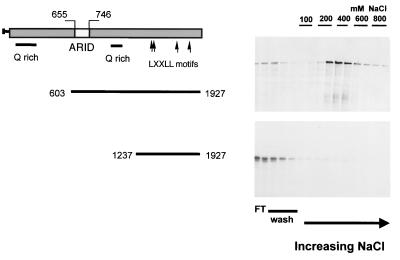
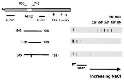
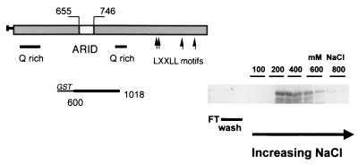
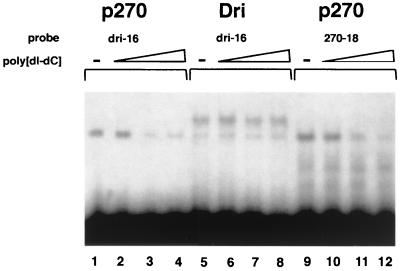
p270 is an integral member of human SWI-SNF complexes, first identified through its shared antigenic specificity with p300 and CREB binding protein. The deduced amino acid sequence of p270 reported here indicates that it is a member of an evolutionarily conserved family of proteins distinguished by the presence of a DNA binding motif termed ARID (AT-rich interactive domain). The ARID consensus and other structural features are common to both p270 and yeast SWI1, suggesting that p270 is a human counterpart of SWI1. The approximately 100-residue ARID sequence is present in a series of proteins strongly implicated in the regulation of cell growth, development, and tissue-specific gene expression. Although about a dozen ARID proteins can be identified from database searches, to date, only Bright (a regulator of B-cell-specific gene expression), dead ringer (a Drosophila melanogaster gene product required for normal development), and MRF-2 (which represses expression from the cytomegalovirus enhancer) have been analyzed directly in regard to their DNA binding properties. Each binds preferentially to AT-rich sites. In contrast, p270 shows no sequence preference in its DNA binding activity, thereby demonstrating that AT-rich binding is not an intrinsic property of ARID domains and that ARID family proteins may be involved in a wider range of DNA interactions.


Figures

FIG. 1
FIG. 1
Sequences of peptides obtained from the microsequencing of p270. Left column, sequences of four peptides derived from the peptide sequencing of gel-purified p270 isolated from human cells; middle column, sequence of the multiple-antigen peptide (map) that was used to raise the p270-specific antibodies used in the cloning; right column, sequences of the corresponding regions deduced from the p270 cDNA clone, with the position number of the initial residue indicated for reference to Fig. 3. X represents uncertainty in reading the protein sequence. The underlining indicates a single discrepancy between the sequence obtained by direct sequencing and that obtained by sequencing the cDNA.
FIG. 2
FIG. 2
Hybridization of p270 cDNA sequence with an ∼8.0-kb RNA band in multitissue Northern blots. Blots were probed with a 2.5-kb cDNA corresponding to the 3′ terminus of the p270 mRNA. Molecular sizes (left) are according to the measurements provided by the supplier. INT, intestine; PBL, peripheral blood lymphocytes; SKEL. MUSC., skeletal muscle.
FIG. 3
FIG. 3
DNA sequence and deduced amino acid sequence of human p270. The nucleotide sequence of the human p270 cDNA is represented along with the deduced amino acid sequence of the product of the 5,781-bp open reading frame. The four LXXLL motifs and the ARID motif are underlined. Two Q-rich regions are present, spanning residues 45 to 253 and 969 to 1072. The sequence also includes a series of seven CAG repeats (34) indicated by underlining beginning at nucleotide 2907.
FIG. 3
FIG. 3
DNA sequence and deduced amino acid sequence of human p270. The nucleotide sequence of the human p270 cDNA is represented along with the deduced amino acid sequence of the product of the 5,781-bp open reading frame. The four LXXLL motifs and the ARID motif are underlined. Two Q-rich regions are present, spanning residues 45 to 253 and 969 to 1072. The sequence also includes a series of seven CAG repeats (34) indicated by underlining beginning at nucleotide 2907.
FIG. 4
FIG. 4
A comparison of the ARID regions from p270 with those of other ARID-containing proteins. The boundaries of the ARID region were originally defined in Bright and DRI (9, 11). These sequences are aligned here together with others more recently added to the database. Black boxes, identical residues; gray boxes, conservative differences. The consensus sequence consists of 36 highly conserved residues with an approximately 95-residue stretch. All sequences were obtained from public protein databases. Accession numbers are as follows, eyelid, AF053091; SWI1, P09547; Bright, U60335; DRIL1, U88047; DRI, U62542; RBP 1, P29374; RBP 2, P29375; MRF-1, S27962; MRF-2, S27963; smcx, P41229; jumonji, U57592; Arabidopsis thaliana open reading frame (ORF) product f2202.20, AAC83071; C. elegans ORF products c01g8 and t23d8, U80438 and Z81128, respectively.
FIG. 5
FIG. 5
Alignment of p270 and SWI1. p270 and SWI1 have an overall similarity of structure, in that they both contain ARID regions and they both have multiple LXXLL motifs (which are implicated in binding to nuclear hormone receptors), as well as Q-rich regions, which are often associated with transactivation regions. SWI1 has an unusual asparagine- and threonine-rich stretch near the N terminus; we do not yet know if this feature also occurs in p270. Other than these common features, p270 and SWI1 do not show direct sequence homology.
FIG. 6
FIG. 6
DNA binding activity in p270. Reticulocyte lysate-translated 35S-labeled p270 peptides were applied to native DNA cellulose columns. The columns were washed with loading buffer and then with increasing salt concentrations. Aliquots of the flowthrough (FT), wash, and eluted fractions were analyzed on SDS gels and visualized by autoradiography. A p270 peptide encompassing residues 603 to 1927 and containing an intact ARID region was retained on the column (upper panel). A related peptide, encompassing residues 1237 to 1927, with the ARID domain deleted, passed through the column and was recovered primarily in the flowthrough and wash fractions (lower panel). The sequences encompassed by the p270 peptides are indicated schematically to the left of the gels.
FIG. 7
FIG. 7
ARID-dependent DNA binding activity in p270. Reticulocyte lysate-translated 35S-labeled p270 peptides were applied to native DNA cellulose columns and eluted as described for Fig. 6. Aliquots of the flowthrough (FT), wash, and eluted fractions were analyzed in a protein filter binding assay and visualized by autoradiography. The sequences encompassed by the p270 peptides are indicated schematically to the left of the gels. The parallel lines in the bar corresponding to the peptide comprising residues 523 to 1281 represent the two single-residue substitutions at positions 715 and 738. The elution profiles were quantified by phosphorimaging. With the p270543–1018 peptide (upper panel), 31.3% of the total counts per minute were recovered in the flowthrough and wash fractions, while 45.4% eluted in the 200 to 600 mM salt fractions. With peptide p270678–1018 (middle panel), 95.3% of the total counts per minute were recovered in the flowthrough and wash fractions while 2.28% eluted in the 200 to 600 mM salt fractions. With peptide p270523–1281WY (lower panel), 76.2% of the total counts per minute were recovered in the flowthrough and wash fractions while 16.7% eluted in the 200 to 600 mM salt fractions.
FIG. 8
FIG. 8
DNA binding activity of a GST-p270 fusion peptide. GST fusion protein GSTp270600–1018 was passed over a DNA cellulose column and collected in a series of increasing salt fractions as described for Fig. 6. Aliquots of each fraction were separated on SDS-polyacrylamide gels. The electrophoresed proteins were transferred to nitrocellulose blots, probed with GST-specific antibodies, and visualized by chemiluminescence. The fusion protein eluted primarily in the 400 mM salt fractions. The sequences encompassed by the fusion peptide are indicated schematically to the left of the gel.
FIG. 9
FIG. 9
Sequences of oligonucleotides bound by p270. Shown are sequences bound by the p270 fusion protein after six cycles of amplification and selection. The overall percentage of AT residues is 47.9%. The sequences were analyzed by the Wisconsin GCG PileUp program, but no common motifs were detected.
FIG. 10
FIG. 10
p270 shows no sequence preference in an EMSA. The DNA binding activity of p270 (lanes 1 to 4 and 9 to 12) and DRI (lanes 5 to 8) was assayed by gel shift using 32P-labeled oligonucleotide probes p270.18 (lanes 1 to 4) and dri.16 (lanes 5 to 12). Competition assays included unlabeled poly(dI-dC) at a molar excess of approximately 20:1 (lanes 2, 6, and 10), 140:1 (lanes 3, 7, and 11), and 600:1 (lanes 4, 8, and 12).

References

    1. Agulnik A I, Bishop C E, Lerner J L, Agulnik S I, Solovyev V V. Analysis of mutation rates in the SMCY/SMCX genes shows that mammalian evolution is male driven. Mamm Genome. 1997;8:134–138. - PubMed
    1. Altschul S F, Boguski M S, Gish W, Wootton J C. Issues in searching molecular sequence databases. Nat Genet. 1994;6:119–129. - PubMed
    1. Altschul S F, Gish W, Miller W, Myers E W, Lippman D J. Basic local alignment search tool. J Mol Biol. 1990;215:403–410. - PubMed
    1. Chiba H, Muramatsu M, Nomoto A, Kato H. Two human homologues of Saccharomyces cerevisiae SWI2/SNF2 and Drosophila brahma are transcriptional coactivators cooperating with the estrogen receptor and the retinoic acid receptor. Nucleic Acids Res. 1994;22:1815–1820. - PMC - PubMed
    1. Dallas P B, Yaciuk P, Moran E. Characterization of monoclonal antibodies raised against p300: both p300 and CBP are present in intracellular TBP complexes. J Virol. 1997;71:1726–1731. - PMC - PubMed

Publication types