. 2000 May;20(9):3234-44.
doi: 10.1128/MCB.20.9.3234-3244.2000.

Cid1, a fission yeast protein required for S-M checkpoint control when DNA polymerase delta or epsilon is inactivated

Affiliations

Cid1, a fission yeast protein required for S-M checkpoint control when DNA polymerase delta or epsilon is inactivated

S W Wang et al. Mol Cell Biol. 2000 May.

Abstract

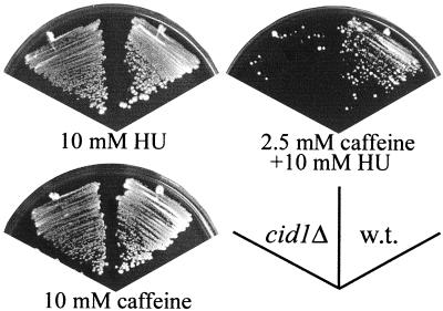
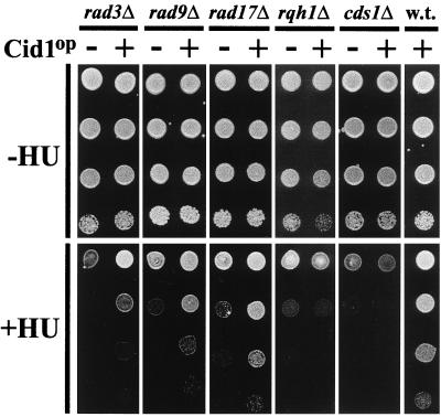
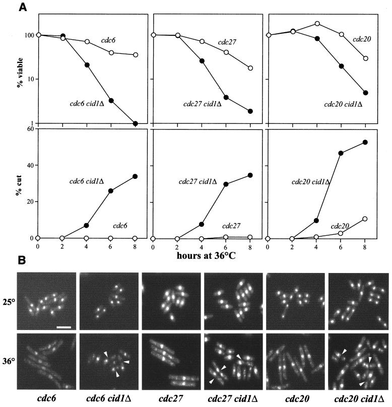
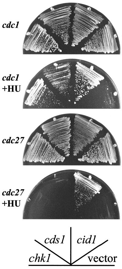
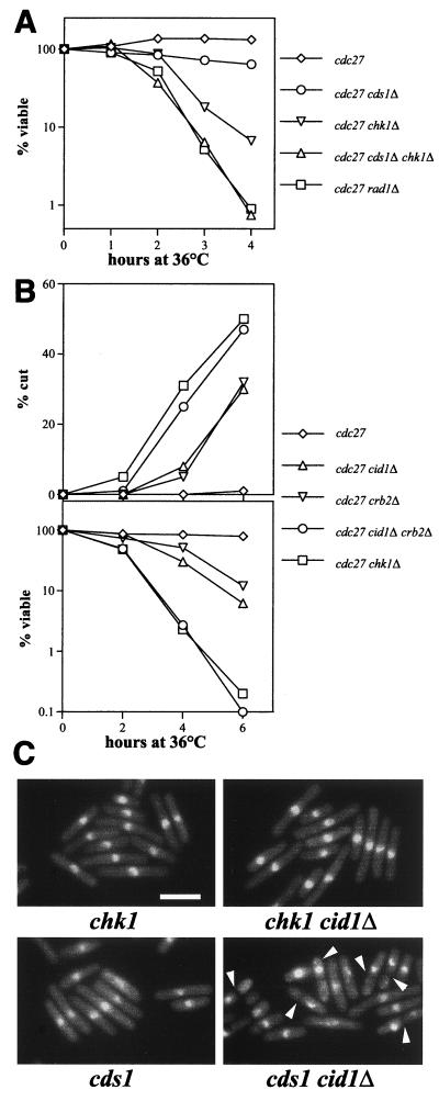
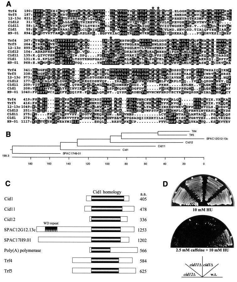
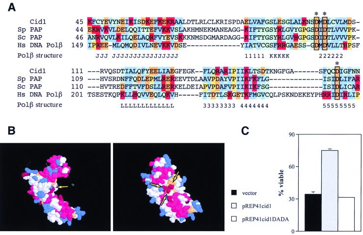
The S-M checkpoint is an intracellular signaling pathway that ensures that mitosis is not initiated in cells undergoing DNA replication. We identified cid1, a novel fission yeast gene, through its ability when overexpressed to confer specific resistance to a combination of hydroxyurea, which inhibits DNA replication, and caffeine, which overrides the S-M checkpoint. Cid1 overexpression also partially suppressed the hydroxyurea sensitivity characteristic of DNA polymerase delta mutants and mutants defective in the "checkpoint Rad" pathway. Cid1 is a member of a family of putative nucleotidyltransferases including budding yeast Trf4 and Trf5, and mutation of amino acid residues predicted to be essential for this activity resulted in loss of Cid1 function in vivo. Two additional Cid1-like proteins play similar but nonredundant checkpoint-signaling roles in fission yeast. Cells lacking Cid1 were found to be viable but specifically sensitive to the combination of hydroxyurea and caffeine and to be S-M checkpoint defective in the absence of Cds1. Genetic data suggest that Cid1 acts in association with Crb2/Rhp9 and through the checkpoint-signaling kinase Chk1 to inhibit unscheduled mitosis specifically when DNA polymerase delta or epsilon is inhibited.


Figures

FIG. 1
FIG. 1
Deletion of cid1 confers sensitivity specifically to the combination of HU and low-dose caffeine. Fission yeast strains HM123 (wild type [w.t.]) and cid1Δ were streaked onto YPD agar plates containing 10 mM HU, 10 mM HU plus 2.5 mM caffeine, or 10 mM caffeine, as indicated. The plates were photographed after 5 to 7 days of incubation at 30°C.
FIG. 2
FIG. 2
Overexpression of Cid1 partially suppresses the HU sensitivity of checkpoint rad mutants. Cells of strains rad3Δ, rad9Δ, rad17Δ, rqh1Δ, cds1Δ, and HM123 (wild type [w.t.]) transformed with either pREP1 (−) or pREP1cid1 (+) were plated at 10-fold serial dilutions either onto minimal agar supplemented with adenine (−HU) or onto the same agar additionally supplemented with 2 mM (rad3Δ, rad9Δ and rad17Δ) or 5 mM (rqh1Δ and cds1Δ) HU (+HU). The plates were photographed after 5 days of incubation at 30°C.
FIG. 3
FIG. 3
Deletion of cid1 causes loss of checkpoint integrity when Pol δ or ɛ is inhibited. t.s. Pol δ (cdc6, cdc27) and Pol ɛ (cdc20) strains and the respective double mutants with cid1Δ, as indicated, were grown in liquid culture to mid-logarithmic phase at 25°C and shifted to 36°C, the restrictive temperature. (A) Samples of 500 cells taken at the indicated times after the shift to 36°C were plated in duplicate onto YPD agar and incubated at 25°C. After 4 days of growth, viability (top panels) was scored as a percentage of the number of colonies formed by the sample taken at time zero. Samples taken at the same time points were fixed, DAPI stained, and examined by fluorescence microscopy. The percentage of each sample exhibiting the cut phenotype (bottom panels) was scored by counting a total of at least 200 cells for each time point. (B) Representative fields of DAPI-stained cells of the indicated strains grown at 25°C (top panels) or 6 h after the shift to 36°C (bottom panels). Cut cells are indicated (arrowheads). Bar, 10 μm.
FIG. 4
FIG. 4
Cid1 overexpression partially suppresses the HU sensitivity of cdc1-P13 and cdc27-P11 mutants. cdc1 or cdc27 strains transformed with pREP1cid1, pREP1cds1, pREPchk1, or an “empty” vector (pREP1) as indicated were streaked onto YPD plates or plates containing 5 mM (cdc1) or 10 mM (cdc27) HU. The plates were photographed after 5 days of growth at 30°C.
FIG. 5
FIG. 5
Checkpoint integrity is dependent on Cid1, Crb2, and Chk1 when Cdc27 is inactivated or when cds1-deleted cells are exposed to HU. (A and B) The indicated strains were shifted from 25 to 36°C, and cell viability (A) or viability and the percentage of cells exhibiting the cut phenotype (B) were determined as described in the legend to Fig. 3. (C) The indicated strains were grown to mid-log phase in YPD medium at 30°C prior to the addition of HU to 10 mM. Cells were fixed, DAPI stained, and examined by fluorescence microscopy. Representative fields of cells fixed 5 h after HU addition are shown, and cut cells are indicated (arrowheads). Bar, 10 μm.
FIG. 6
FIG. 6
Cid1 belongs to a novel protein family in S. pombe. (A) Alignment of the predicted protein sequences of Cid1 and related proteins in S. pombe and S. cerevisiae. Only the region of significant similarity to Cid1 (approximately 300 amino acid residues) is shown in each case, with amino acid residue numbers given on the left. 12-13c denotes the predicted product of SPAC12G12-13c, and H9-01 denotes the predicted product of SPAC17H9-01. Amino acid residues found at the same position in three or more of the aligned sequences are shaded in black, and conservative substitutions are highlighted in grey. The conserved aspartate triad residues are indicated by asterisks. (B) Cladogram showing the relationship between Cid1 family members in S. pombe and the Trf4 and Trf5 proteins of S. cerevisiae. The length of each pair of branches represents the distance between sequence pairs. Units indicate the number of substitution events. (C) Schematic representation of the overall structural similarity between Cid1, Cid11, Cid12, SPAC12G12.13c, SPAC17H9.01, and poly(A) polymerase from S. pombe and Trf4 and Trf5 from S. cerevisiae. The extent of the region of significant similarity between these proteins is indicated by the shaded area, and the location of the seven tandem WD repeats in SPAC12G12.13c is also shown. (D) Deletion of any one of the smaller cid1-related genes results in sensitivity to HU in the presence of low-dose caffeine. Strains HM123 (wild type [w.t.]), cid1Δ, cid11Δ, and cid12Δ were streaked as indicated onto YPD agar containing 10 mM HU or 2.5 mM caffeine plus 10 mM HU. The plates were photographed after 7 days of incubation at 30°C.
FIG. 7
FIG. 7
Cid1 is a putative nucleotidyltransferase. (A) Alignment of the amino acid sequences of Cid1, poly(A) polymerases from S. pombe (Sp) and S. cerevisiae (Sc), and human Pol β in the region of the aspartate triad (boxed) involved in catalysis [based on the poly(A) polymerase alignment of Martin and Keller [28]). The positions of α-helices (J, K, L) and β-strands (1 to 5) in the corresponding region of the crystal structure of rat Pol β (40) are indicated below the alignment. Human and rat Pol β sequences differ at only one position in the region shown. Amino acid residue groups are color coded as follows: blue, hydrophobic; red, positively charged; orange, negatively charged; green, polar; yellow, proline. (B) Predicted structure for Cid1 generated by superimposition of Cid1 amino acid side chains 1 to 236 on the Cα structure of rat Pol β (40). Two alternative views of the structure, generated using RasMol, are shown, with the clustered aspartate triad indicated (arrows). (C) Mutation of the aspartate triad of Cid1 leads to loss of checkpoint-signaling function. The t.s. cdc27 cid1Δ strain was transformed with pREP41cid1, pREP41cid1DADA, or an “empty” vector (pREP41). Transformants were grown for 16 h in EMM2 medium lacking thiamine before being shifted to 36°C for 6 h; viability was measured as described in the legend to Fig. 3.

Similar articles

Cited by

References

    1. Al-Khodairy F, Carr A M. DNA repair mutants defining G2 checkpoint pathways in Schizosaccharomyces pombe. EMBO J. 1992;11:1343–1350. - PMC - PubMed
    1. Al-Khodairy F, Fotou E, Sheldrick K S, Griffiths D J, Lehmann A R, Carr A M. Identification and characterization of new elements involved in checkpoint and feedback controls in fission yeast. Mol Biol Cell. 1994;5:147–160. - PMC - PubMed
    1. Altschul S F, Madden T L, Schäffer A A, Zhang J, Zhang Z, Miller W, Lipman D J. Gapped BLAST and PSI-BLAST: a new generation of protein database search programs. Nucleic Acids Res. 1997;25:3389–3402. - PMC - PubMed
    1. Aravind L, Koonin E V. DNA polymerase β-like nucleotidyltransferase superfamily: identification of three new families, classification and evolutionary history. Nucleic Acids Res. 1999;27:1609–1618. - PMC - PubMed
    1. Bahler J, Wu J Q, Longtine M S, Shah N G, McKenzie III A, Steever A B, Wach A, Philippsen P, Pringle J R. Heterologous modules for efficient and versatile PCR-based gene targeting in Schizosaccharomyces pombe. Yeast. 1998;14:943–951. - PubMed

Publication types

MeSH terms