. 2000 May;20(9):3245-55.
doi: 10.1128/MCB.20.9.3245-3255.2000.

A novel family of cell wall-related proteins regulated differently during the yeast life cycle

Affiliations

A novel family of cell wall-related proteins regulated differently during the yeast life cycle

J M Rodríguez-Peña et al. Mol Cell Biol. 2000 May.

Abstract

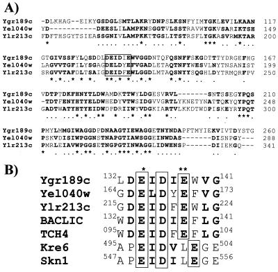
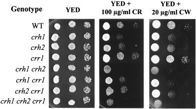
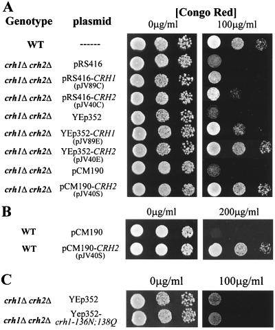
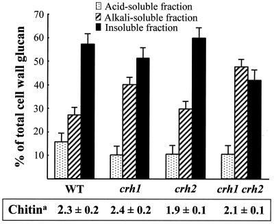
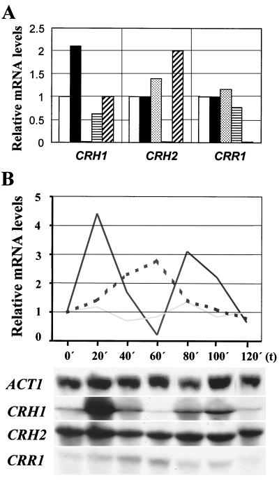
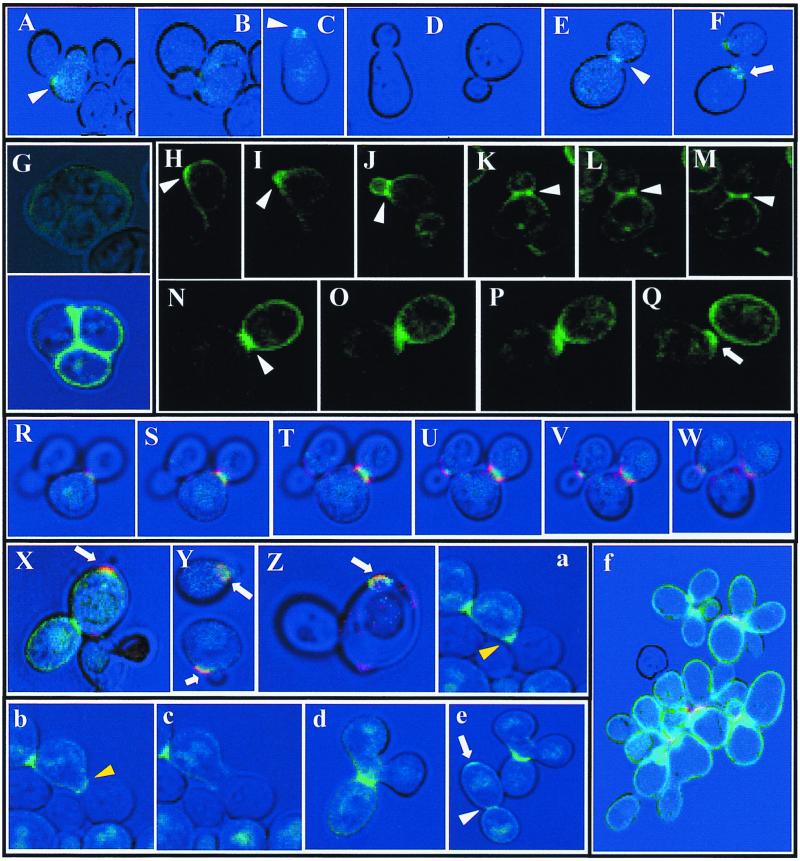
The Saccharomyces cerevisiae Ygr189c, Yel040w, and Ylr213c gene products show significant homologies among themselves and with various bacterial beta-glucanases and eukaryotic endotransglycosidases. Deletion of the corresponding genes, either individually or in combination, did not produce a lethal phenotype. However, the removal of YGR189c and YEL040w, but not YLR213c, caused additive sensitivity to compounds that interfere with cell wall construction, such as Congo red and Calcofluor White, and overexpression of YEL040w led to resistance to these compounds. These genes were renamed CRH1 and CRH2, respectively, for Congo red hypersensitive. By site-directed mutagenesis we found that the putative glycosidase domain of CRH1 was critical for its function in complementing hypersensitivity to the inhibitors. The involvement of CRH1 and CRH2 in the development of cell wall architecture was clearly shown, since the alkali-soluble glucan fraction in the crh1Delta crh2Delta strain was almost twice the level in the wild-type. Interestingly, the three genes were subject to different patterns of transcriptional regulation. CRH1 and YLR213c (renamed CRR1, for CRH related) were found to be cell cycle regulated and also expressed under sporulation conditions, whereas CRH2 expression did not vary during the mitotic cycle. Crh1 and Crh2 are localized at the cell surface, particularly in chitin-rich areas. Consistent with the observed expression patterns, Crh1-green fluorescent protein was found at the incipient bud site, around the septum area in later stages of budding, and in ascospore envelopes. Crh2 was found to localize mainly at the bud neck throughout the whole budding cycle, in mating projections and zygotes, but not in ascospores. These data suggest that the members of this family of putative glycosidases might exert a common role in cell wall organization at different stages of the yeast life cycle.


Figures

FIG. 1
FIG. 1
(A) Amino acid alignment of homologous Ygr189c, Yel040w, and Ylr213c sequences. Identical and conserved residues are boldfaced and are indicated by asterisks and dots, respectively. The putative catalytic domain similar to that of the 1,3-1,4-β-glucanases from Bacillus licheniformis is boxed. (B) Amino acid alignment of homologous sequences within the proposed catalytic domain of bacterial endo-β-1,3-1,4-glucanases (48), corresponding to Ygr189c, Yel040w, Ylr213c, Kre6, and Skn1 from S. cerevisiae, B. licheniformis 1,3-1,4-β-glucanase (BACLIC), and A. thaliana xyloglucan endotransglycosidase (TCH4). A single asterisk identifies the proposed catalytic nucleophile (18, 21), and two asterisks indicate the position of the proposed general acid-base residue (49). Amino acids that are identical in all the sequences are boxed. Boldface indicates residues of conserved properties with respect to the B. licheniformis domain in more than three sequences. Numbers correspond to the amino acid positions in the protein sequences.
FIG. 2
FIG. 2
Sensitivities to Congo red and Calcofluor white of strains bearing all the possible combinations of deletions of the CRH1, CRH2, and CRR1 genes. Cells were grown in YED, and 1/10 dilution series of each strain were spotted onto YED plates containing the indicated amounts of Congo red and Calcofluor white.
FIG. 3
FIG. 3
(A) Complementation studies on a crh1 crh2 deletion strain growing on YED plates containing Congo red at the amounts indicated and transformed with various plasmids harboring the wild-type (WT) CRH1 or CRH2 gene in a pRS416-based centromeric vector, a YEp352-based episomic vector, or the pCM190-based overexpression vector (see Table 2). (B) Resistance to Congo red induced by overexpression of CRH2 in a wild-type background. (C) A mutant crh1-136N,138Q allele does not complement the sensitivity to Congo red in the crh1Δ crh2Δ strain.
FIG. 4
FIG. 4
Determination of the alkali-insoluble–acid-soluble, alkali-soluble, and alkali-insoluble–acid-insoluble fractions of cell wall glucan in wild-type, crh1Δ, crh2Δ, and crh1Δ crh2Δ cells. The whole glucan fraction that remained after protease treatment of purified cell walls was considered 100%. a, the chitin content for each strain, in micrograms of glucosamine per milligram (dry weight) of cells, is given.
FIG. 5
FIG. 5
Northern blot analysis for the study of the expression of the CRH1, CRH2, and CRR1 genes. (A) Relative mRNA levels for CRH1, CRH2, and CRR1. mRNA expression levels in a wild-type background when cells were grown in glucose-rich medium (open bars) were assigned a value of 1 for each hybridization assay. The amount of RNA was normalized to that of ACT1 mRNA (a constitutively expressed gene in S. cerevisiae). mRNA levels for each gene in a wild-type background when cells were grown in galactose-based YEG medium (solid bars), in a crh1Δ strain grown in YED (stippled bars), in a crh2Δ background (horizontally striped bars), and in a crr1Δ strain (diagonally striped bars) are also shown. (B) Northern blot analysis of CRH1, CRH2, and CRR1 expression during the cell cycle in synchronized haploid cells after release from pheromone-induced arrest (see Materials and Methods). A relative value of 1 was assigned to RNA levels for each transcript at time 0. The exposure times for each film were as follows: 2 h for ACT1, overnight for CRH1 and CRH2, and 10 days for CRR1. Solid line, CRH1; shaded line, CRH2; dashed line, CRR1.
FIG. 6
FIG. 6
Cellular localization by confocal microscopy of Crh1-GFP and Crh2-GFP. (A through F) Crh1-GFP visualized in crh1 cells bearing the pJV89G plasmid during vegetative growth. (A and B) Images of the same cells 45 min apart. The fluorescent patch in panel A marks the site of bud emergence. (C through F) Cells from the same transformant after release from mating pheromone-induced arrest in different stages of the budding cycle: cells with incipient budding (C), with medium-sized buds (D), with large buds (E), and after separation (F). (G) A double homozygous crh1 crh2 diploid strain transformed with pJV89G and allowed to sporulate, showing Crh1-GFP localization in ascospores. (H through Q) Time lapse localization of Crh2-GFP in crh2-deleted cells bearing the pJV40G plasmid. The pictures show a sequence of images taken every 10 min on a growing bud from the stage of bud emergence (H and I), every 20 min during bud growth (J through M), and during the stages of cytokinesis and cell separation at intervals of 10 min, except for the last one (30 min) (N through Q). (R through W) Confocal analysis of six slides (0.5-μm width) of budding cells expressing Chr2-GFP. Red color, chitin-rich areas detected by WGA-TRITC staining; green color, localization of the protein. (X through Y) Colocalization of Crh2-GFP and WGA-TRITC-stained chitin at the bud scar that remains from the last cytokinetic event in both diploid (X) and haploid (Y) backgrounds. (Z) Colocalization of Crh1-GFP and WGA-TRITC-stained chitin at the bud scar. (a through e) crh2Δ strain transformed with pJV40G in the process of mating to untransformed cells of the opposite mating type. The sequence in panels a through c shows the localization of Crh2-GFP in the mating projection and throughout the process of cell fusion (time lapse between pictures, 15 min). In panel d, a mature zygote is shown, with Crh2p marking the cytokinesis site. The image in panel e depicts the same zygote after 90 min, when it had developed a second diploid bud. A residual Crh2-GFP mark is apparent at the birth site of the first diploid, showing the bipolar polarity pattern characteristic of diploids. (f) Localization of Crh2-GFP in a chs2::LEU2 crh2Δ background. White arrowheads indicate accumulation of GFP fusion proteins at incipient bud sites and bud necks. Yellow arrowheads indicate accumulation of fluorescence at the shmoo. Arrows point to GFP-Crh localization at bud scars.

References

    1. Arroyo J, Garcia-Gonzalez M, Garcia-Saez M I, Sanchez M, Nombela C. DNA sequence analysis of a 23,002 bp DNA fragment of the right arm of Saccharomyces cerevisiae chromosome VII. Yeast. 1997;13:357–363. - PubMed
    1. Ausubel F M, Brent R, Kingston R E, Moore D D, Seidman J G, Smith J A, Struhl K. Current protocols in molecular biology. New York, N.Y: Greene Publishing Associates and Wiley Interscience; 1993.
    1. Cabib E, Roberts R, Bowers B. Synthesis of the yeast cell wall and its regulation. Annu Rev Biochem. 1982;51:763–793. - PubMed
    1. Campbell P, Braam J. Co- and/or post-translational modifications are critical for TCH4 XET activity. Plant J. 1998;15:553–561. - PubMed
    1. Caro L H P, Tettelin H, Vossen J H, Ram A F J, Van Den Ende H, Klis F M. In silicio identification of glycosyl-phosphatidylinositol-anchored plasma-membrane and cell wall proteins of Saccharomyces cerevisiae. Yeast. 1997;13:1477–1489. - PubMed

Publication types

MeSH terms