. 2000 May;20(9):3256-65.
doi: 10.1128/MCB.20.9.3256-3265.2000.

Dual control of muscle cell survival by distinct growth factor-regulated signaling pathways

Affiliations

Dual control of muscle cell survival by distinct growth factor-regulated signaling pathways

M A Lawlor et al. Mol Cell Biol. 2000 May.

Abstract

In addition to their ability to stimulate cell proliferation, polypeptide growth factors are able to maintain cell survival under conditions that otherwise lead to apoptotic death. Growth factors control cell viability through regulation of critical intracellular signal transduction pathways. We previously characterized C2 muscle cell lines that lacked endogenous expression of insulin-like growth factor II (IGF-II). These cells did not differentiate but underwent apoptotic death in low-serum differentiation medium. Death could be prevented by IGF analogues that activated the IGF-I receptor or by unrelated growth factors such as platelet-derived growth factor BB (PDGF-BB). Here we analyze the signaling pathways involved in growth factor-mediated myoblast survival. PDGF treatment caused sustained activation of extracellular-regulated kinases 1 and 2 (ERK1 and -2), while IGF-I only transiently induced these enzymes. Transient transfection of a constitutively active Mek1, a specific upstream activator of ERKs, maintained myoblast viability in the absence of growth factors, while inhibition of Mek1 by the drug UO126 blocked PDGF-mediated but not IGF-stimulated survival. Although both growth factors activated phosphatidylinositol 3-kinase (PI3-kinase) to similar extents, only IGF-I treatment led to sustained stimulation of its downstream kinase, Akt. Transient transfection of a constitutively active PI3-kinase or an inducible Akt promoted myoblast viability in the absence of growth factors, while inhibition of PI3-kinase activity by the drug LY294002 selectively blocked IGF- but not PDGF-mediated muscle cell survival. In aggregate, these observations demonstrate that distinct growth factor-regulated signaling pathways independently control myoblast survival. Since IGF action also stimulates muscle differentiation, these results suggest a means to regulate myogenesis through selective manipulation of different signal transduction pathways.


Figures
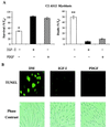
FIG. 1
FIG. 1
IGF-I and PDGF promote myoblast survival. (A) Cell counts of living (left) or dead (right) C2AS12 myoblasts after a 24-h incubation in DM without or with R3IGF-I (2 nM) or PDGF-BB (0.4 nM). The asterisk at the left indicates that significantly fewer cells survived (P < 0.003; mean ± SE of three experiments) in untreated than in growth factor-treated cells; asterisks at the right indicate that significantly more cells died (P < 0.0005; mean ± SE of three experiments). (B) Results of TUNEL assays of C2AS12 cells 24 h after incubation in DM or in DM supplemented with R3IGF-I (2 nM) or PDGF-BB (0.4 nM). The lower panel shows phase-contrast views of the cells. (C) Results of cell counts of living (left) or dead (right) C2 myoblasts after a 24-h incubation in DM without or with R3IGF-I (0.4 nM) or PDGF-BB (0.3 nM). The asterisk at the left panel indicates that significantly fewer cells survived (P < 0.0001; mean ± SE of three experiments) in untreated than in growth factor-treated cells; asterisks at the right indicate that significantly more cells died (P < 0.0003; mean ± SE of three experiments).
FIG. 1
FIG. 1
IGF-I and PDGF promote myoblast survival. (A) Cell counts of living (left) or dead (right) C2AS12 myoblasts after a 24-h incubation in DM without or with R3IGF-I (2 nM) or PDGF-BB (0.4 nM). The asterisk at the left indicates that significantly fewer cells survived (P < 0.003; mean ± SE of three experiments) in untreated than in growth factor-treated cells; asterisks at the right indicate that significantly more cells died (P < 0.0005; mean ± SE of three experiments). (B) Results of TUNEL assays of C2AS12 cells 24 h after incubation in DM or in DM supplemented with R3IGF-I (2 nM) or PDGF-BB (0.4 nM). The lower panel shows phase-contrast views of the cells. (C) Results of cell counts of living (left) or dead (right) C2 myoblasts after a 24-h incubation in DM without or with R3IGF-I (0.4 nM) or PDGF-BB (0.3 nM). The asterisk at the left panel indicates that significantly fewer cells survived (P < 0.0001; mean ± SE of three experiments) in untreated than in growth factor-treated cells; asterisks at the right indicate that significantly more cells died (P < 0.0003; mean ± SE of three experiments).
FIG. 2
FIG. 2
PDGF treatment leads to sustained stimulation of ERK1 and -2. (A) Representative immunoblot using a phospho-specific ERK1 and -2 antibody and whole cell protein extracts from C2AS12 cells treated with either PDGF (0.4 nM) or R3IGF-I (2 nM) for the indicated times (top) and the same samples after incubation with an antibody to total ERKs (bottom). Results are representative of three experiments. (B) Results of an in vitro ERK kinase assay performed with immunoprecipitates from cells incubated with either PDGF (0.4 nM) or R3IGF-I (2 nM) for the indicated times as described in Materials and Methods (top) and results from three experiments (mean ± SE) presented in graphical form relative to values measured after 5 min of PDGF treatment, set arbitrarily to 100% (bottom).
FIG. 3
FIG. 3
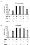
Treatment with an inhibitor of Mek1 attenuates PDGF-mediated muscle cell survival. (A) Confluent C2AS12 myoblasts were treated with PDGF (0.4 nM) or R3IGF-I (2 nM) for 18 h, followed by a 6-h incubation with either growth factor plus UO126 (10 μM). Cell counts of viable myoblasts were performed at 24 h. The graph presents results of four experiments, each performed in duplicate (mean ± SE). The asterisks indicate that significantly fewer cells survived after incubation with PDGF plus UO126 than in the other treatment groups (∗, P < 0.0005; ∗∗, P < 0.00005; #, P < 0.005). (B) Confluent C2 cells were treated with PDGF (0.3 nM) or R3IGF-I (0.4 nM) for 18 h, followed by a 6-h incubation with either growth factor plus UO126 (10 μM). Cell counts of viable myoblasts were performed at 24 h. The graph presents results of four experiments, each performed in duplicate (mean ± SE). The asterisks indicate that significantly fewer cells survived after incubation with PDGF plus UO126 than in the other treatment groups (∗, P < 0.0005; ∗∗, P < 0.02).
FIG. 4
FIG. 4
Forced expression of active Mek1 maintains myoblast survival. (A) C2AS12 myoblasts were transiently transfected with recombinant expression plasmids encoding either a constitutively active Mek1 (Mek1*) or wild-type enzyme (Mek1WT). Cell counts of transfected myoblasts were performed 24 h after incubation in DM or in DM supplemented with PDGF (0.4 nM). Results show the mean ± SE of five experiments, each performed in duplicate. There was significantly less survival of cells transfected with the Mek1WT plasmid than of cells transfected with Mek1* (P < 0.0005). (B) Representative immunoblot of an in vitro ERK kinase assay performed with protein extracts of cells transfected with recombinant expression plasmids for either Mek1* or Mek1WT. The graph shows results (mean ± SE) of three experiments performed in duplicate. There was significantly less activity in cells transfected with the Mek1WT plasmid than in cells transfected with Mek1* (P < 0.006).
FIG. 5
FIG. 5
Reversal of muscle cell survival by the Mek1 inhibitor UO126. Transfected myoblasts were incubated in DM or in DM supplemented with PDGF (0.4 nM) for 18 h, followed by addition of UO126 (10 μM) for 6 h. Cell counts of viable myoblasts were performed at 24 h. Results are expressed as the mean ± SE of four independent experiments, each performed in duplicate. The asterisk denotes that survival was significantly less in myoblasts treated with UO126 than in untreated cells (P < 0.0003).
FIG. 6
FIG. 6
Both IGF-I and PDGF stimulate PI3-kinase activity. (A) Autoradiograph showing results of an in vitro PI3-kinase assay using cell extracts from confluent myoblasts incubated in DM without or with R3IGF-I (2 nM) or PDGF-BB (0.4 nM) for 5 min, performed as described in Materials and Methods. The location of the origin and the migration of the 3-phosphorylated products are indicated on the right. (B) Results of time course studies measuring PI3-kinase enzymatic activity after treatment of muscle cells with R3IGF-I (2 nM) or PDGF-BB (0.4 nM). Values on the y axis represent arbitrary densitometric units. Results are representative of three experiments.
FIG. 7
FIG. 7
A PI3-kinase inhibitor blocks IGF-mediated myoblast survival. (A) C2AS12 cells were incubated in DM or in DM supplemented with PDGF (0.4 nM) or R3IGF-I (2 nM) for 18 h, followed by addition of LY294002 (30 μM) for 6 h. Cell counts of viable myoblasts were performed at 24 h. Results are expressed as the mean ± SE of three independent experiments, each performed in duplicate. The asterisks denote that survival was significantly less in myoblasts treated with R3IGF-I plus LY294002 than in the other groups (∗, P < 0.0002; ∗∗, P < 0.00002; #, P < 0.003). (B) C2 myoblasts were incubated in DM or in DM supplemented with PDGF (0.3 nM) or R3IGF-I (0.4 nM) for 18 h, followed by addition of LY294002 (30 μM) for 6 h. Cell counts of viable myoblasts were performed at 24 h. Results are expressed as the mean ± SE of three independent experiments, each performed in duplicate. The asterisks denote that survival was significantly less in myoblasts treated with R3IGF-I plus LY294002 than in the other groups (∗, P < 0.0005, ∗∗, P < 0.003).
FIG. 8
FIG. 8
Forced expression of active PI3-kinase maintains myoblast survival. (A) C2AS12 myoblasts were transiently transfected with recombinant expression plasmids encoding either a constitutively active PI3-kinase (p110*) or an inactive enzyme (p110Δkin). Counts of transfected cells were performed 24 h after incubation in DM or in DM supplemented with R3IGF-I (2 nM). Results show the mean ± SE of four experiments, each performed in duplicate. There was significantly less survival of cells transfected with the p110Δkin plasmid than of cells transfected with p110* (P < 0.004). (B) Immunoblot of protein extracts from Cos7 cells transiently transfected with an expression plasmid for EGFP, p110*, or p110Δkin, using an antibody for the Myc epitope tag present in the latter two fusion genes.
FIG. 9
FIG. 9
Reversal of muscle cell survival by the PI3-kinase inhibitor LY294002. Transfected C2AS12 myoblasts were incubated in DM or in DM supplemented with R3IGF-I (2 nM) for 18 h, followed by addition of LY294002 (30 μM) for 6 h. Cell counts of viable myoblasts were performed at 24 h. Results are expressed as the mean ± SE of three independent experiments, each performed in duplicate. The asterisks denote that survival was significantly less in myoblasts treated with LY294002 than in untreated cells (∗, P < 0.003; ∗∗, P < 0.0002).
FIG. 10
FIG. 10
IGF treatment leads to sustained activation of Akt. (A) Representative immunoblot using a phospho-specific Akt antibody and whole cell protein extracts from C2AS12 cells treated with either R3IGF-I (2 nM) or PDGF (0.4 nM) for the indicated times (top) and the same blot after being stripped and incubated with an antibody for total Akt (bottom). Results are representative of three experiments. (B) Results of a representative in vitro Akt kinase assay performed with immunoprecipitates from cells incubated with either R3IGF-I (2 nM) or PDGF (0.4 nM) for the indicated times as described outlined in Materials and Methods (top) and results from three experiments (mean ± SE) presented in graphical form (bottom). Values on the y axis represent arbitrary densitometric units.
FIG. 11
FIG. 11
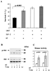
Akt promotes muscle cell survival. (A) C2AS12 myoblasts were transiently transfected with a recombinant expression plasmid encoding an inducible Akt (iAkt). Cell counts of transfected myoblasts were performed 24 h after incubation in DM or in DM supplemented with HT, R3IGF-I (2 nM), or both agents. Results are expressed as the mean ± SE of four experiments, each performed in duplicate. There was significantly greater survival in transfected cells treated with HT than in untreated cells (P < 0.003). (B) Representative immunoblot using an antibody specific for phospho-Akt and cell extracts from transfected myoblasts incubated without or with HT for the indicated times (top) and the same blot after being stripped and incubated with an antibody to total Akt (bottom). (C) Results of in vitro Akt kinase assays (mean ± SE of four experiments) performed with immunoprecipitates from transfected cells incubated without or with HT for the indicated times as described in Materials and Methods. Values on the y axis represent arbitrary densitometric units. Significantly more Akt enzymatic activity was measured after treatment with HT at both time points, as indicated on the graph.

Similar articles

Cited by

References

    1. Alessi D R, Andjelkovic M, Caudwell B, Cron P, Morrice N, Cohen P, Hemmings B A. Mechanism of activation of protein kinase B by insulin and IGF-1. EMBO J. 1996;15:6541–6551. - PMC - PubMed
    1. Anderson C N G, Tolkovsky A M. A role for MAPK/ERK in sympathetic neuron survival: protection against a p53-dependent, JNK-independent induction of apoptosis by cytosine arabinoside. J Neurosci. 1999;19:664–673. - PMC - PubMed
    1. Antonetti D A, Algenstaedt P, Kahn C R. Insulin receptor substrate 1 binds two novel splice variants of the regulatory subunit of phosphatidylinositol 3-kinase in muscle and brain. Mol Cell Biol. 1996;16:2195–2203. - PMC - PubMed
    1. Barton-Davis E R, Shoturma D I, Musaro A, Rosenthal N, Sweeney H L, Kurabayashi M, Dutta S, Kedes L. Viral mediated expression of insulin-like growth factor I blocks the aging-related loss of skeletal muscle function. Proc Natl Acad Sci USA. 1998;95:15603–15607. - PMC - PubMed
    1. Baserga R, Resnicoff M, D'Ambrosio C, Valentinis B. The role of the IGF-I receptor in apoptosis. Vitam Horm. 1997;53:65–98. - PubMed

Publication types

MeSH terms

Substances

LinkOut - more resources