. 2000 Apr 25;97(9):4742-7.
doi: 10.1073/pnas.080063497.

Muir-Torre-like syndrome in Fhit-deficient mice

Affiliations

Muir-Torre-like syndrome in Fhit-deficient mice

L Y Fong et al. Proc Natl Acad Sci U S A. .

Abstract

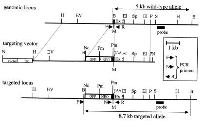
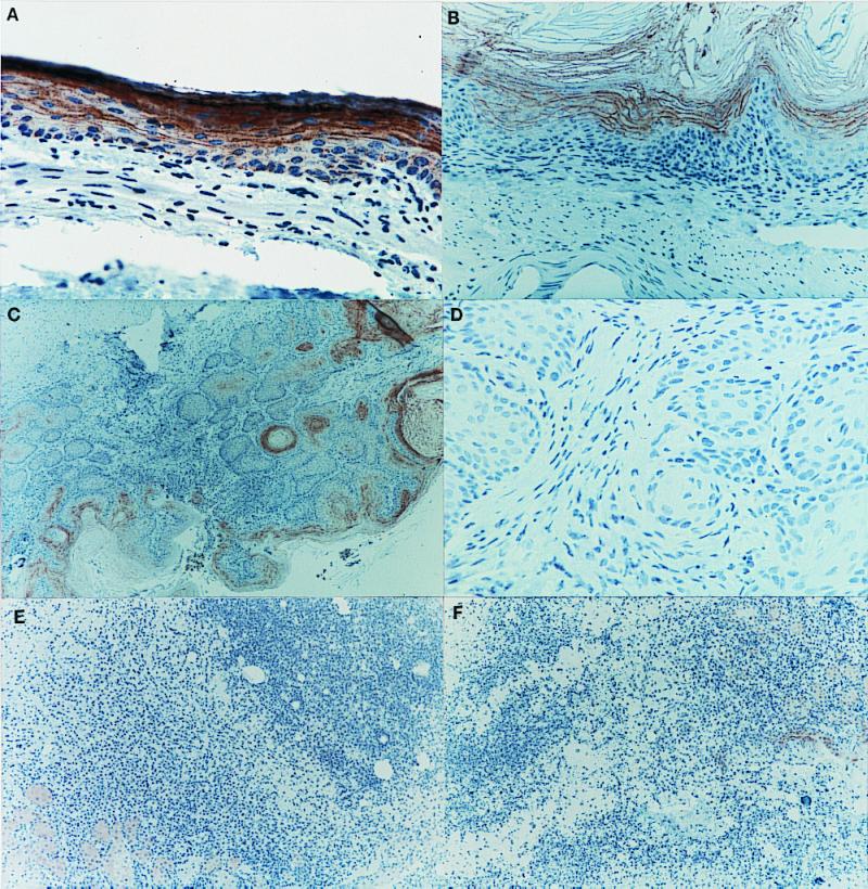
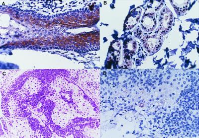
To investigate the role of the Fhit gene in carcinogen induction of neoplasia, we have inactivated one Fhit allele in mouse embryonic stem cells and produced (129/SvJ x C57BL/6J) F(1) mice with a Fhit allele inactivated (+/-). Fhit +/+ and +/- mice were treated intragastrically with nitrosomethylbenzylamine and observed for 10 wk posttreatment. A total of 25% of the +/+ mice developed adenoma or papilloma of the forestomach, whereas 100% of the +/- mice developed multiple tumors that were a mixture of adenomas, squamous papillomas, invasive carcinomas of the forestomach, as well as tumors of sebaceous glands. The visceral and sebaceous tumors, which lacked Fhit protein, were similar to those characteristic of Muir-Torre familial cancer syndrome.


Figures

Figure 1
Figure 1
Murine Fhit genomic locus, targeting and screening strategy. The top line represents the Fhit genomic locus surrounding exon 5. The middle line depicts the targeting vector with a 6.6-kb HindIII (H)–PstI (P) fragment with a termination codon introduced into exon 5. The targeted locus after homologous recombination is shown at the bottom with the probe used for Southern blot screening of ES colony and progeny DNA after BamHI (B) cleavage. Positions of the primers used for PCR amplification of progeny DNA, to identify wild-type (F,R) and targeted (N,R) alleles, are shown. Restriction enzyme sites are shown for EcoRV (EV), EcoRI (E1), SphI (Sp), SacI (S), NotI (N), NcoI (Nc), and Pme1 (Pm). The 5′→3′ sequences of the three primers F, R, and N are, respectively: CTTGAATCTAGGCTGCATTCTAGCGAG, GATTCCTTGCTTACCTTTTGGGGATGG, and TGGGCTCTATGGCTTCTGAGGC. The first reaction product is a wild-type fragment of ≈450 bp containing exon 5; the second product is a mutant fragment of ≈280 bp spanning from the Neo selection gene to intron 5. PCR conditions were: denaturation 94°C, 30 s; annealing 62°C, 30 s; elongation 72°C, 30 s; 35 cycles.
Figure 2
Figure 2
Absence of Fhit protein in the Fhit −/− mice. Lysates from tissues of Fhit −/− mice were tested for expression of Fhit by immunoblot analysis of mouse tissue lysates: lane 1, Fhit +/+ lung; lane 2, +/+ liver; lane 3, +/+ kidney; lane 4, Fhit −/− liver; lane 5, −/− kidney.
Figure 3
Figure 3
Immunohistochemical detection of Fhit expression. (A) Fhit expression in normal esophageal epithelium (×200) of Fhit +/+ mouse 23 at 10 wk post NMBA; the brown chromogen represents the Fhit protein. (B) Lack of Fhit expression in a squamous papilloma of the forestomach (×200) in Fhit +/− mouse 33 at 10 wk post NMBA; (C) Absence of Fhit expression in a squamous papilloma of the junction (×200) in Fhit +/+ mouse 25 at 10 wk post NMBA; (D) Lack of Fhit expression in an invasive squamous carcinoma of the forestomach (×100) in Fhit +/− mouse 31 at 10 wk post NMBA; (E) Lack of Fhit expression in a sebaceous tumor (×100) in Fhit +/− mouse 27 at 10 wk post NMBA; (F) Absence of Fhit protein in a sebaceous tumor (×100) in Fhit +/− mouse 21 at 10 wk post NMBA.
Figure 4
Figure 4
Immunohistochemical detection of human Fhit in MTS tumors. (A) Fhit expression in normal hair follicle (×200); note that dense keratin horn shows nonspecific staining; (B) Fhit expression in normal sebaceous gland (×200); (C) H&E staining of an MTS case 1 sebaceous tumor; (D) Lack of Fhit expression in most cells of the case 1 sebaceous tumor.
Figure 5
Figure 5
Integrity of Fhit loci in murine tumors. DNA from tails and sebaceous tumors was cleaved with XbaI, electrophoresed, transferred to a membrane, and hybridized to a 32P-labeled full-length Fhit cDNA probe. Fhit exons are indicated on the left; the asterisk indicates the inactivated Fhit exon 5. Lanes 1, 3, and 4 contained DNAs from sebaceous tumors from Fhit +/− mice 21, 27, and 31; lane 2 contained DNA from the tail of Fhit +/+ mouse 25, and lane 5 contained DNA from a Swiss mouse 3T3 cell line, which exhibits a variant-sized exon 3 (obscured by another fragment) because of a polymorphism. The Fhit +/+ and +/− mice are B6129F1s, which exhibit two different alleles of exon 8. (Right) The agarose gel before blotting of the digested DNAs to the membrane; this gel illustrates that amounts of DNA loaded in individual lanes varied from ≈1 μg (lane 4) to ≈10 μg (lane 2).
Figure 6
Figure 6
Assessment of MSI in tumors. DNA templates from mouse and human tumors and controls were amplified by using primers flanking microsatellite alleles. Labeled amplified products were run on PAGE gels, dried, and exposed. The D6Mit59, D19Mit36, and D17Mit123 panels represent murine alleles amplified from Fhit +/− mouse 27 forestomach tumor (lane 1), Fhit +/+ mouse 25 forestomach tumor (lane 2), Fhit +/+ mouse 25 tail (lane 3), Fhit +/− mouse 21 sebaceous tumor (lane 4), Fhit +/− mouse 27 sebaceous tumor (lane 5), Fhit +/− mouse 27 second sebaceous tumor (lane 6), Fhit +/− mouse 31 sebaceous tumor (lane 7), K1735 mouse melanoma cell line (lane 8), NP3 mouse cell line (lane 9), negative control (no DNA) (lane 10). No MSI was observed in the mouse tumors for the three markers shown. The D18S35 and D3S1295 panels represent germline and tumor DNA from a human MTS case: DNA from peripheral blood lymphocytes (lane 1), DNA from sebaceous tumor 185 from the same individual (lane 2), lymphocyte DNA (lane 3), and DNA from sebaceous tumors 185 (lane 4) and 9029 (lane 5) from the same individual. These sebaceous tumors showed MSI at each allele successfully amplified.

References

    1. Yunis J J, Soreng A L. Science. 1984;226:1199–1204. - PubMed
    1. Ohta M, Inoue H, Cotticelli M G, Kastury K, Baffa R, Palazzo J, Siprashvili Z, Mori M, McCue P, Druck T, et al. Cell. 1996;84:587–597. - PubMed
    1. Sozzi G, Veronese M L, Negrini M, Baffa R, Cotticelli M G, Inoue H, Tornielli S, Pilotti S, DeGregorio L, Pastorino V, et al. Cell. 1996;85:17–26. - PubMed
    1. Hendricks D T, Taylor R, Reed M, Birrer M J. Cancer Res. 1997;57:2112–2115. - PubMed
    1. Greenspan D L, Connolly D C, Wu R, Lei R Y, Vogelstein J T C, Kim Y-T, Mok J E, Munoz N, Bosch X, Shah K, Cho K R. Cancer Res. 1997;57:4692–4698. - PubMed

Publication types

MeSH terms