. 2000 Apr;122(4):1099-108.
doi: 10.1104/pp.122.4.1099.

The expression of small heat shock proteins in seeds responds to discrete developmental signals and suggests a general protective role in desiccation tolerance

Affiliations

The expression of small heat shock proteins in seeds responds to discrete developmental signals and suggests a general protective role in desiccation tolerance

N Wehmeyer et al. Plant Physiol. 2000 Apr.

Abstract

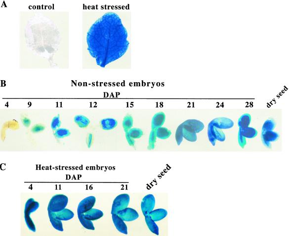
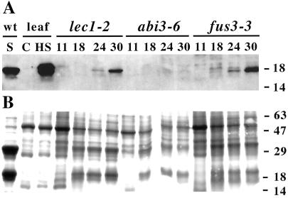
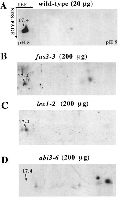
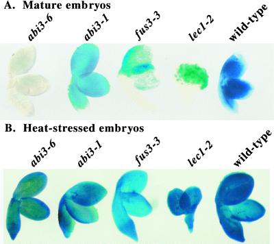
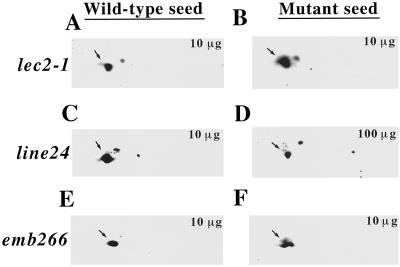
To learn more about the function and regulation of small heat shock proteins (sHSPs) during seed development, we studied sHSP expression in wild-type and seed maturation mutants of Arabidopsis by western analysis and using an HSP17.4 promoter-driven beta-glucuronidase (GUS) reporter gene in transgenic plants. In the absence of stress, GUS activity increases during development until the entire embryo is stained before desiccation. Heat-stressed embryos stained for GUS at all stages, including early stages that showed no detectable HSP17. 4::GUS activity without heat. Examination of HSP17.4 expression in seeds of the transcriptional activator mutants abi3-6, fus3-3 (AIMS no. CS8014/N8014), and lec1-2 (AIMS no. CS2922/N2922) showed that protein and HSP17.4::GUS activity were highly reduced in fus3-3 and lec1-2 and undetectable in abi3-6 seeds. In contrast, heat-stressed abi3-6, fus3-3, and lec1-2 seeds stained for GUS activity throughout the embryo. These data indicate that there is distinct developmental and stress regulation of HSP17.4, and imply that ABI3 activates HSP17.4 transcription during development. Quantitation of sHSP protein in desiccation-intolerant seeds of abi3-6, fus3-3, lec1-2, and line24 showed that all had <2% of wild-type HSP17.4 levels. In contrast, the desiccation-tolerant but embryo-defective mutants emb266 (AIMS no. CS3049/N3049) and lec2-1 (AIMS no. CS2728/N2728) had wild-type levels of HSP17.4. These data correlate a reduction in sHSPs with desiccation intolerance and suggest that sHSPs have a general protective role throughout the seed.


Figures

Figure 1
Figure 1
HSP17.4::GUS activity in heat-stressed leaves, developing embryos, and heat-stressed embryos of Arabidopsis. A, Arabidopsis leaves (T3 generation) non-stressed (control) or heat-stressed. B, Arabidopsis embryos (T3) stained for GUS activity during seed maturation. Seeds were collected at the designated times, and embryos were dissected away from the seed coat for staining. All non-stressed samples were stained with X-gluc for 4 h at room temperature, and heat-stressed leaves were stained for 30 min at 37°C. C, Arabidopsis seeds were heat-stressed for 4 h at 38°C, embryos were dissected from the seed coat at the corresponding time, and stained with X-gluc for 30 min at 37°C (4 h at room temperature yielded the same results).
Figure 2
Figure 2
HSP17.4 accumulation in mutants of the seed transcriptional activators LEC1, ABI3, and FUS3. Seeds were collected from abi3-6, lec1-2, and fus3-3 homozygous mutant plants during seed development at the indicated times (nos. above the lanes in DAP). For comparison, heat-stressed leaf (HS), control leaf (C), and wild-type seed (S) samples were also analyzed. A, Total seed proteins separated by SDS-PAGE and analyzed by western blotting with anti-HSP17.6 antibodies. B, Total protein profile of samples in A visualized on separate gels by staining with Coomassie Blue. Ten micrograms of total seed protein was loaded in each lane. Molecular mass markers are indicated on the right (in kD).
Figure 3
Figure 3
HSP17.4 accumulates to <2% of wild-type levels in dry seeds from transcriptional activator mutants (dry seed samples are equivalent to 30 DAP). Total seed proteins from wild type or the indicated mutants were separated by two-dimensional electrophoresis and analyzed by western blotting with HSP17.6 antibodies. A, Wild-type seed proteins (20 μg). B, fus3-3 seed proteins (200 μg). C, lec1-2 seed proteins (200 μg). D, abi3-6 seed proteins (200 μg). Only a portion of the SDS-PAGE is shown, however, there were no other significant cross-reacting polypeptides. The position of HSP17.4 is indicated with an arrow.
Figure 4
Figure 4
Comparison of developmental and heat regulation of HSP17.4::GUS in abi3-6, fus3-3, and lec1-2 mutants. A, Mature (30 DAP) wild-type embryos or homozygous mutant embryos stained 4 h at room temperature for HSP17.4::GUS activity in the absence of heat. B, Embryos stained for GUS activity directly after a heat stress. Mutant embryos represent F3 seeds from the appropriate cross. Embryos were dissected from the seed coat and stained as described in “Materials and Methods.”
Figure 5
Figure 5
HSP17.4 levels are reduced only in desiccation-intolerant seed mutants, not in mutants with other defects in embryogenesis. Heterozygous mutant plants were grown to collect homozygous mutant or phenotypically wild-type seed, as described in “Materials and Methods.” For desiccation-tolerant lec2-1: A, phenotypically wild-type seed; B, homozygous mutant. For desiccation-intolerant line 24: C, phenotypically wild-type seed; D, homozygous mutant. For desiccation-tolerant emb266: E, phenotypically wild-type seed; F, homozygous mutant. Total seed proteins (10 or 100 μg as indicated) were extracted from dry seeds, separated by two-dimensional electrophoresis, blotted to nitrocellulose, and probed with anti-HSP17.6 antibodies. Dry seed samples showed similar results to earlier time points (approximately 21 DAP) as analyzed by one-dimensional electrophoresis.

References

    1. Alamillo J, Almogura C, Bartels D, Jordano J. Constitutive expression of small heat shock proteins in vegetative tissues of the resurrection plant Craterostigma plantagineum. Plant Mol Biol. 1995;29:1093–1099. - PubMed
    1. Almoguera C, Jordano J. Developmental and environmental concurrent expression of sunflower dry-seed stored low-molecular weight heat-shock proteins during late embryogenesis. Plant Mol Biol. 1992;19:781–792. - PubMed
    1. Almoguera C, Prieto-Dapena P, Jordano J. Dual regulation of a heat shock promoter during embryogenesis: stage-dependent role of heat shock elements. Plant J. 1998;13:437–446. - PubMed
    1. Bechtold N, Pelletier G. In planta Agrobacterium-mediated transformation of adult Arabidopsis thaliana plants by vacuum infiltration. Methods Mol Biol. 1998;82:259–266. - PubMed
    1. Bernal-Lugo I, Leopold A. The dynamics of seed mortality. J Exp Bot. 1998;49:1455–1461.

Publication types

MeSH terms

Substances

LinkOut - more resources