. 2000 Apr;122(4):1365-77.
doi: 10.1104/pp.122.4.1365.

Changes in Hechtian strands in cold-hardened cells measured by optical microsurgery

Affiliations

Changes in Hechtian strands in cold-hardened cells measured by optical microsurgery

C S Buer et al. Plant Physiol. 2000 Apr.

Abstract

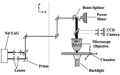
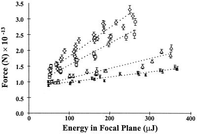
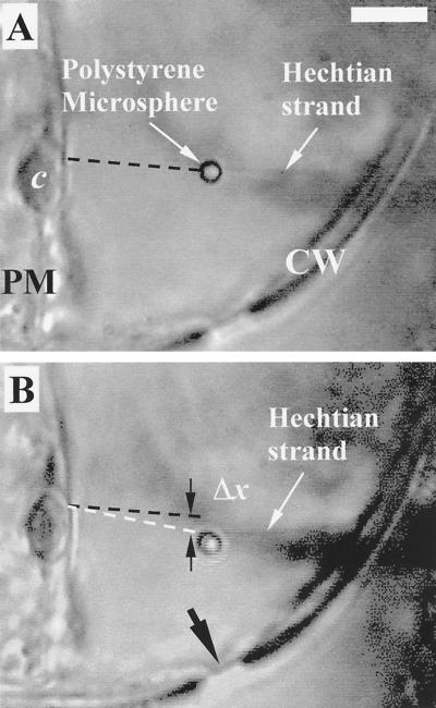
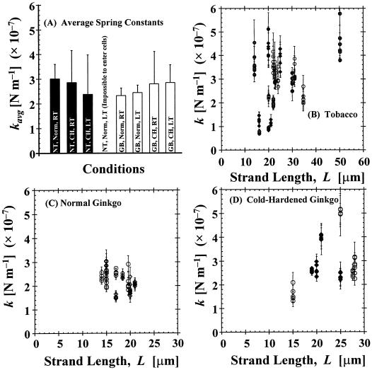
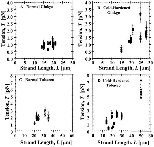
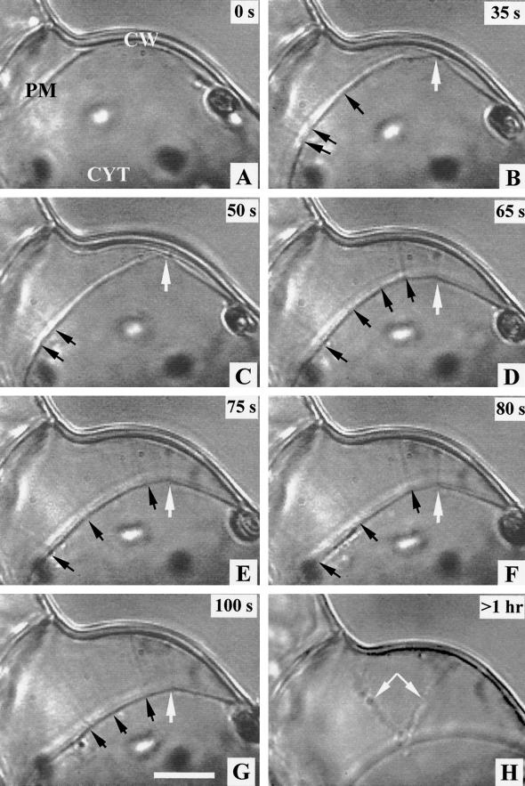
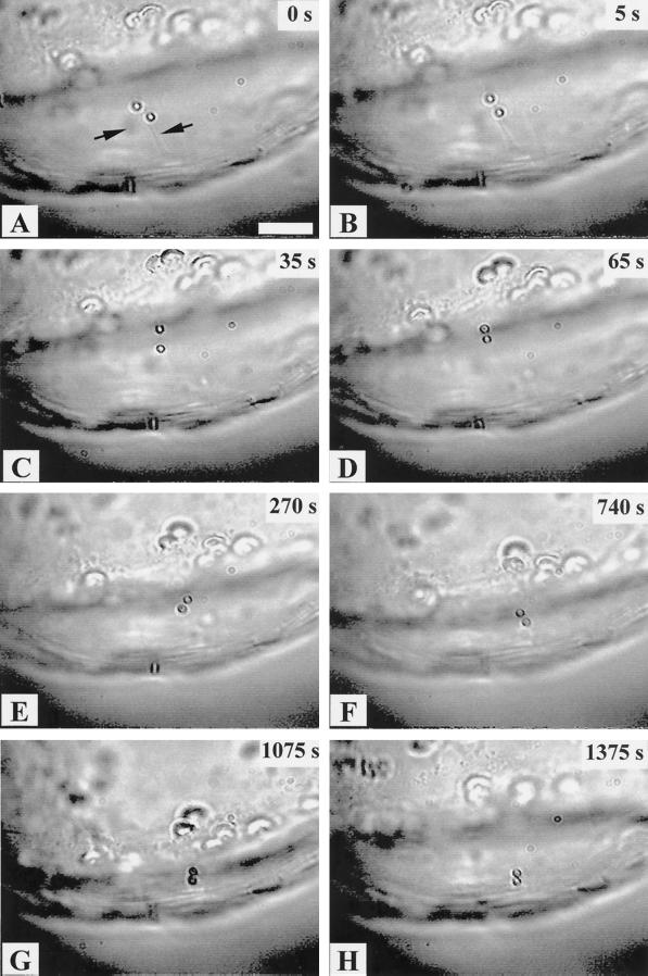
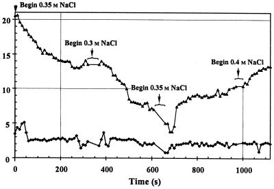
Optical microsurgical techniques were employed to investigate the mechanical properties of Hechtian strands in tobacco (Nicotiana tabacum) and Ginkgo biloba callus cells. Using optical tweezers, a 1. 5-microm diameter microsphere coated with concanavalin A was inserted though an ablated hole in the cell wall of a plasmolyzed cell and attached to a Hechtian strand. By displacing the adhered microsphere from equilibrium using the optical trapping force, the tensions of individual strands were determined. Measurements were made using both normal and cold-hardened cells, and in both cases, tensions were on the order of 10(-12) N. Significant differences were found in the binding strengths of cold-hardened and normal cultured cells. An increased number density of strands in cold-hardened G. biloba compared with normal cultured cells was also observed. Although no Hechtian strands were detected in any Arabidopsis callus cells, strands were present in leaf epidermal cells. Finally, the movement of attached microspheres was monitored along the outside of a strand while cycling the osmotic pressure.


Figures

Figure 1
Figure 1
Schematic of the optical system. A Nd:YAG laser supplies a Q-switched UV beam for ablating a hole through the cell wall and a long-pulsed infrared beam for optical trapping of polystyrene microspheres. The beam is directed to a ×100 microscope objective, and the beam forms a focal spot within a chamber containing callus cells. Backlighting the chamber allows the objective to image the trapping region onto a CCD camera.
Figure 2
Figure 2
Calibration of average trapping force versus energy in the focal plane for 1.5-μm diameter Con A-coated polystyrene microspheres using Stokes' Law following the method of Wright et al. (1994). Error bars are the se of the mean of at least 20 separate trials in 0.4 m NaCl, 10 mm Tris, pH 7.2, 0.01% (w/v) calcofluor, 10 mm CaCl2, and 10 mm MnCl2. ⋄, 20 μm depth; □, 40 μm depth; ▵, 60 μm depth; ×, 80 μm depth.
Figure 3
Figure 3
Videotape frames showing a Hechtian strand (with an attached 1.5-μm diameter polystyrene microsphere) between the cell wall (CW) and plasmalemma (PM) of a plasmolyzed Ginkgo biloba cell. A, Equilibrium position (no displacement) showing the relative position of the strand (dashed black line) with a convenient chloroplast (c). Strand displaced by a distance, Δx (B, dashed white line) compared with the equilibrium position (A). The ablated hole though which the microsphere was manipulated is marked in B (arrow). Bar is 5 μm.
Figure 4
Figure 4
Measured values of the spring constant (k) when a Hechtian strand of length L is transversely displaced from equilibrium. Measurements were made at room temperature (RT) at approximately 20°C (circles) and low temperature (LT) at approximately 10°C (diamonds). Values of k for normal (Norm) RT, ○; cold-hardened (CH), LT, ●, and Norm LT, ♦, for tobacco, (B); Norm G. biloba (GB) RT, ○, and Norm GB LT, ⋄, (C); and cold-hardened G. biloba, RT, ○, and LT, ♦ (D).
Figure 5
Figure 5
Measured values of the tension in Hechtian strands for normal G. biloba (A), cold-hardened G. biloba (B), normal N. tabacum (C), and cold-hardened N. tabacum (D).
Figure 6
Figure 6
Sequence from videotape of a normal cultured N. tabacum callus cell showing Hechtian strand formation during plasmolysis. A, Incipient plasmolysis at 0.3 m NaCl. B through G, Plasmolysis with the gradual introduction of 0.4 m NaCl. Time of sequence is indicated in the upper right corners, and strands are at small arrows. Photograph of the same cell about an hour later showing changes in the strands (H, arrows). An apparent strong attachment point (B–G, large arrow) has disappeared in H. These strands were not used for tension measurements. The apparent depth of field is approximately 1 μm. CYT, Cytoplasm; PM, plasma membrane; and CW, cell wall. Bar is 10 μm.
Figure 7
Figure 7
Sequence from a videotape of a typical deplasmolysis/replasmolysis experiment with 1.5 μm-diameter Con A-coated microspheres attached to the same Hechtian strand (A, arrows). A plot of the microsphere movement for this sequence is shown in Figure 8. Time of the sequence is in the upper right-hand corner of each frame. A through C, 0.4 → 0.35 m NaCl step-gradient; D and E, 0.35 → 0.3 m NaCl; F, 0.3 m, just before introduction of 0.35 m NaCl; G and H, 0.35 → 0.4 m NaCl gradient. Bar is 10 μm.
Figure 8
Figure 8
Measured distances between the centers of the attached microspheres (♦) and the position of the protoplast (with respect to the cell wall) as the cell first deplasmolyzes (0–610 s) and then replasmolyzes (660–1,120 s), corresponding to Figure 7 (▴). Varying the concentration of NaCl in steps controlled the osmotic pressure. Distance measurements have an uncertainty of ±0.5 μm. The mean distance between the microspheres is 2.7 ± 0.7 μm and 2.0 ± 0.4 μm during deplasmolysis and replasmolysis, respectively.

References

    1. Akama K, Shiraishi H, Ohta S, Nakamura K, Okada K, Shimura Y. Efficient transformation of Arabidopsis thaliana: comparison of the efficiencies with various organs, plant ecotypes, and Agrobacterium strains. Plant Cell Rep. 1992;12:7–11. - PubMed
    1. Alberdi M, Corcuera LJ. Cold acclimation in plants. Phytochemistry. 1991;30:3177–3184.
    1. Ashkin A, Dziedzic JM. Internal cell manipulation using infrared traps. Proc Natl Acad Sci USA. 1989;86:7914–7918. - PMC - PubMed
    1. Ashkin A, Dziedzic JM, Bjorkholm JE, Chu S. Observations of a single-beam gradient force optical trap for dielectric particles. Optics Lett. 1986;11:288–290. - PubMed
    1. Attree SM, Sheffield E. Plasmolysis of Pteridium protoplasts: a study using light and scanning-electron microscopy. Planta. 1985;165:151–157. - PubMed

Publication types

LinkOut - more resources