. 2000 Apr 11;97(8):4005-10.
doi: 10.1073/pnas.070052697.

Cadherin interaction probed by atomic force microscopy

Affiliations

Cadherin interaction probed by atomic force microscopy

W Baumgartner et al. Proc Natl Acad Sci U S A. .

Abstract

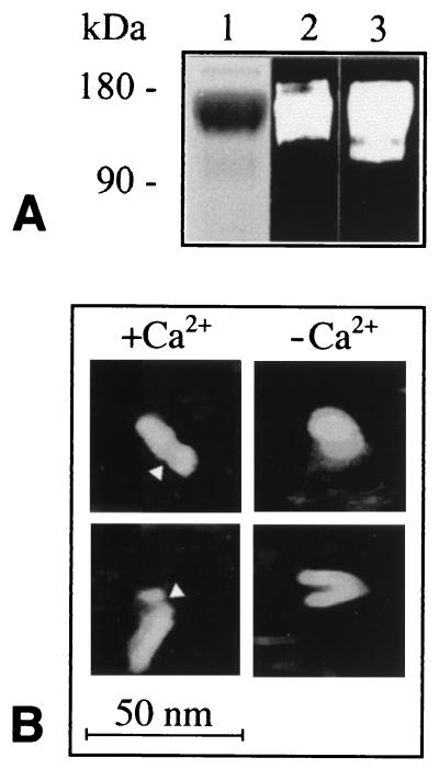
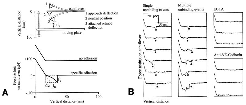
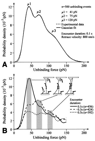
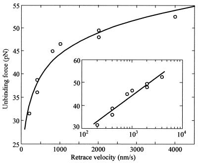
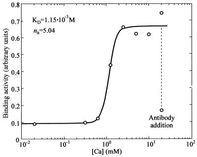
Single molecule atomic force microscopy was used to characterize structure, binding strength (unbinding force), and binding kinetics of a classical cadherin, vascular endothelial (VE)-cadherin, secreted by transfected Chinese hamster ovary cells as cis-dimerized full-length external domain fused to Fc-portion of human IgG. In physiological buffer, the external domain of VE-cadherin dimers is a approximately 20-nm-long rod-shaped molecule that collapses and dissociates into monomers (V-shaped structures) in the absence of Ca(2+). Trans-interaction of dimers is a low-affinity reaction (K(D) = 10(-3)-10(-5) M, k(off) = 1.8 s(-1), k(on) = 10(3)-10(5) M(-1) x s(-1)) with relatively low unbinding force (35-55 pN at retrace velocities of 200-4,000 nm x s(-1)). Higher order unbinding forces, that increase with interaction time, indicate association of cadherins into complexes with cumulative binding strength. These observations favor a model by which the inherently weak unit binding strength and affinity of cadherin trans-interaction requires clustering and cytoskeletal immobilization for amplification. Binding is regulated by low-affinity Ca(2+) binding sites (K(D) = 1.15 mM) with high cooperativity (Hill coefficient of 5.04). Local changes of free extracellular Ca(2+) in the narrow intercellular space may be of physiological importance to facilitate rapid remodeling of intercellular adhesion and communication.


Figures

Figure 1
Figure 1
Purified VE-cadherin-Fc characterized by SDS/PAGE and immunoblotting (A) and by AFM imaging (B). (A) SDS/PAGE was stained with Coomassie blue (1), and immunoblots were probed with VE-cadherin antibody (mAb 11D4.1) (2) and antibody against human IgG (3). Electrophoretic mobility (160–180 kDa) corresponds exactly to the calculated molecular weight of the chimeric dimer. (B) AFM images of hydrated VE-cadherin-Fc adsorbed to mica and scanned in the presence and absence of Ca2+ in isotonic buffer. Note elongated rod-like structure in Ca2+ and globular to v-shaped morphology in the absence of Ca2+. Arrowheads point to constrictions assumed to mark the boundary between the Fc-portion and the cadherin moiety.
Figure 2
Figure 2
General working principles of force measurements (A) and examples of force distance recordings (B). (A) Chimeric VE-cadherin-Fc is covalently attached by PEG linkers to plate and cantilever tip of the AFM. Cadherins bound to tip and plate by flexible PEG linkers are capable of free equilibrium diffusion and interaction with opposing cadherins during tip-to-plate encounter. Molecules are brought into binding contact by upward movement of the plate. During downmovement the force required for unbinding (fu) is proportional to deflection to the cantilever (Δz) and its known spring constant. The total length of the stretched PEG-adhesion dimer is defined as unbinding length (lu). (B) Force-distance curves obtained at a retrace velocity of 800 nm·s−1 and 0.1 s encounter duration in buffer A containing 2 mM Ca2+. Examples of single unbinding events and multiple unbinding events are shown. Asterisks mark unbinding events taken for statistical evaluation. Unbinding events were completely abolished by addition of EGTA (5 mM) and antibody to VE-cadherin external domain.
Figure 3
Figure 3
Frequency distribution of unbinding forces between tip- and plate-attached PEG/VE-cadherin-Fc measured at retrace velocities of 800 nm·s−1 and various tip-to-plate encounter intervals. Frequency distribution is expressed as probability density function (n = number of unbinding events). (A) Data shown fit to three Gaussian distributions with peak values (μ1–μ3) at about 40, 75, and 120 pN. (B) Dependency of the frequency of μ1–μ3 on encounter duration indicates a diffusion-limited reaction underlying multimerization of tip- and plate-bound cadherins. Synchronous dirsuption of independent bonds is highly unlikely (see Discussion).
Figure 4
Figure 4
Dependence of unbinding force of the first peak (corresponding to μ1 in Fig. 3) on retrace velocity plotted in the inset on a logarithmic scale. Each point represents the average value of at least 300 unbinding events measured at a given retrace velocity. The solid line is a numerical fit of the data to modified Bell's expression (τ(fu) = τ0 exp(−lr fu/kBT), where τ0 is the lifetime of unstressed bonds, lr the unbinding width between adhering cadherins, kB Boltzman's constant, T is the absolute temperature, and fu is the unbinding force. The fit yields for τ0 ≈ 0.55 s and for lr ≈ 0.59 nm.
Figure 5
Figure 5
Ca2+-dependence of binding activity between tip- and plate-attached PEG/VE-cadherin-Fc at various Ca2+ concentrations. All measurements were obtained with an individual experimental set-up with stepwise increase of the Ca2+ concentration by fluid exchange. Binding activity is defined as area between the force distance curve and the neutral line. Each point is the average of at least 400 force distance cycles. Note pronounced cooperativity with a Hill coefficient of 5.04 and an apparent KD = 1.15 10−3 M. Addition of antibody to VE-cadherin-Fc at 20 mM Ca2+ caused immediate and almost complete inhibition of binding.

References

    1. Gumbiner G M. Cell. 1996;84:345–357. - PubMed
    1. Takeichi M. Curr Opin Cell Biol. 1995;7:619–627. - PubMed
    1. Lampugnani M G, Dejana E. Curr Opin Cell Biol. 1997;9:674–682. - PubMed
    1. Chothia C, Jones E Y. Annu Rev Biochem. 1997;66:823–862. - PubMed
    1. Shapiro L, Fannon A M, Kwong P D, Thomposn A, Lehmann M S, Grubel G, Legrand J F, Als-Nielsen J, Colman D R, Hendrickson W A. Nature (London) 1995;374:327–337. - PubMed

Publication types

LinkOut - more resources