. 2000 Apr 11;97(8):4380-5.
doi: 10.1073/pnas.070056497.

Involvement of multiple intracellular release channels in calcium sparks of skeletal muscle

Affiliations

Involvement of multiple intracellular release channels in calcium sparks of skeletal muscle

A González et al. Proc Natl Acad Sci U S A. .

Abstract

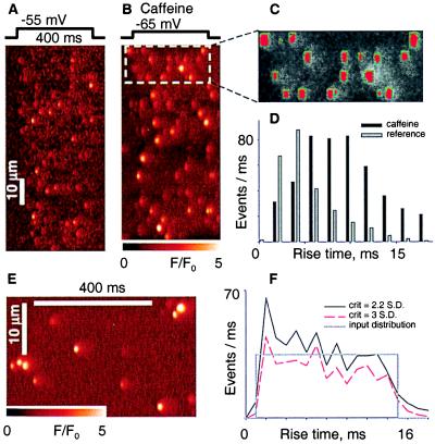
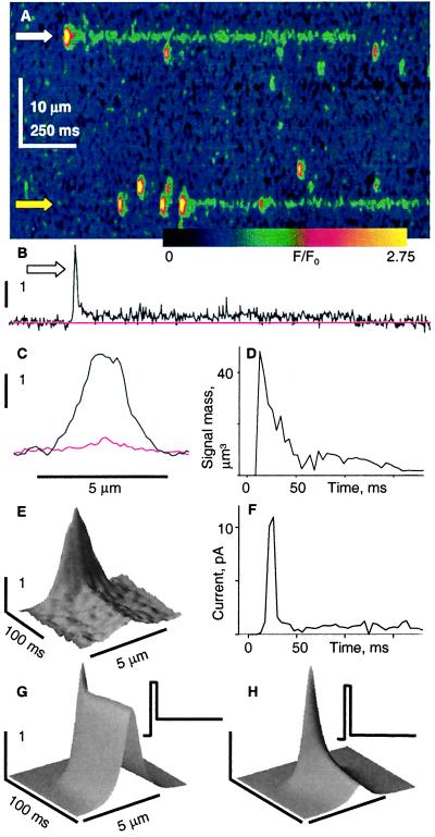
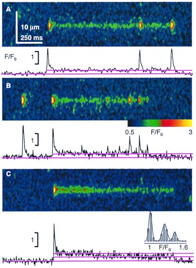
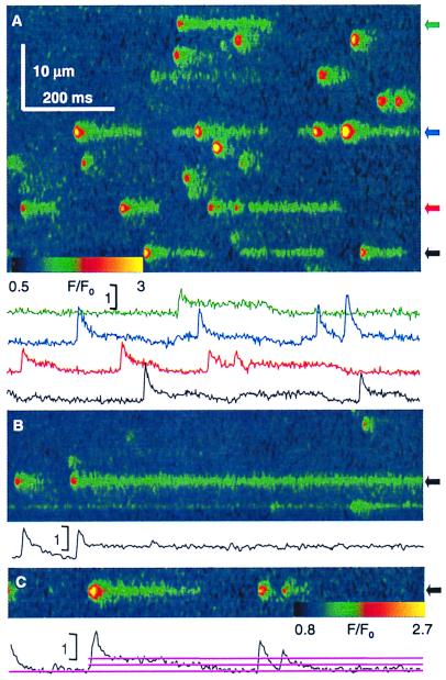
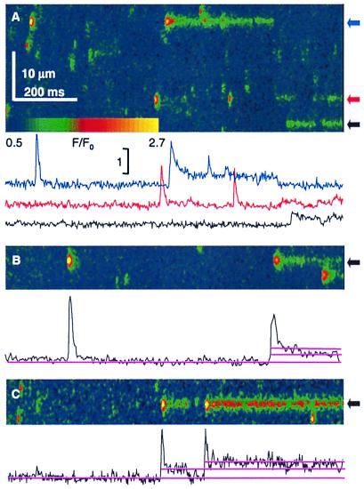
In many types of muscle, intracellular Ca(2+) release for contraction consists of brief Ca(2+) sparks. Whether these result from the opening of one or many channels in the sarcoplasmic reticulum is not known. Examining massive numbers of sparks from frog skeletal muscle and evaluating their Ca(2+) release current, we provide evidence that they are generated by multiple channels. A mode is demonstrated in the distribution of spark rise times in the presence of the channel activator caffeine. This finding contradicts expectations for single channels evolving reversibly, but not for channels in a group, which collectively could give rise to a stereotyped spark. The release channel agonists imperatoxin A, ryanodine, and bastadin 10 elicit fluorescence events that start with a spark, then decay to steady levels roughly proportional to the unitary conductances of 35%, 50%, and 100% that the agonists, respectively, promote in bilayer experiments. This correspondence indicates that the steady phase is produced by one open channel. Calculated Ca(2+) release current decays 10- to 20-fold from spark to steady phase, which requires that six or more channels be open during the spark.


Figures

Figure 1
Figure 1
The distribution of spark rise times. (A) Line-scan image of a fiber held at −90 mV and pulsed to −55 mV as indicated. Fluorescence F normalized to the initial fluorescence F0. (B) The same fiber as in A exposed to 1 mM caffeine externally. (C) The detector's performance: rectangles mark the sides of regions, in red, where F/F0 exceeded 3 SD of the fluorescence in neighboring, nonspark areas (12). (D) Histograms of rise times for 514 events in reference and 938 in caffeine. Cell identifier 0315a. (E) Portion of an image generated placing simulated sparks and Gaussian noise at random positions in an array dimensioned as a line-scan image. Sparks were simulated with source openings lasting between 1 and 15 ms, and seven of each were placed in every image. Sparks were scaled to reproduce off-focus effects and other properties of experimental images (details in Materials and Methods). (F) Rise time histograms of events detected in five simulated images using different threshold criteria. In gray, input distribution, constant between 1 and 15 ms.
Figure 2
Figure 2
Sparks and Iptx-induced openings. (A) Image of a permeabilized cell, immersed in solution with 100 nM Iptx (a gift from H. Valdivia, University of Wisconsin, Madison). (B) Temporal profile of F/F0 at white arrow. (C) Spatial profile of F/F0 at the peak of the starter event and its average (red) during the long event that follows. Identifier 0416b. (D) Time course of signal mass, volume integral of the normalized fluorescence increase in a 6-μm radius. (E) An average of nine long events selected by their large amplitude, from two cells in which the resting fluorescence was similar (0416b, 0423a). F/F0, time and space represented by coordinates z, x, and y, respectively. (F) Release current, calculated from the average event in C (14, 30). (G) Simulated fluorescence event for a Ca2+ source of initial current 10 pA, lasting 6 ms, followed by a steady current of 3.3 pA (Inset). (H) Simulation where the opening at 10 pA is followed by a steady 0.5-pA current. The source diameter was 0.05 μm. Details and parameter values of the simulation were as described in ref. .
Figure 3
Figure 3
Multiple channels within the same release unit. The graphs plot F/F0 averaged over five spatial pixels (0.7 μm) centered at the starter. (A and B) Toxin-induced events with multiple sparks from the same unit. (A) Repeated large sparks indicate that Ca2+ depletion is not a factor in their termination. (B) Sparks of variable amplitudes, coming from the same unit. (C) Example toxin event that decays in two steps of similar amplitude. (Inset) Amplitude histogram from F/F0 after one smoothing step, with Gaussian fit of means 1.01, 1.28, and 1.49. Identifiers were 0423b (A and C) and 0414a (B).
Figure 4
Figure 4
Sparks and long openings induced by Ry. (A) Normalized line-scan image in a permeabilized fiber immersed 15 min in internal solution with 50 nM Ry. Traces plot F/F0 averaged over a 0.7 μm-wide region. Note the long openings, beginning with a starter or, occasionally, without one (black). (B) After ≈50-min exposure frequency of sparks and amplitude of starters decreases. Very long-lasting events are present. (C) Example event that decays in two steps. Identifiers were 1012a66, 1015a60, and 1025a112.
Figure 5
Figure 5
Events in the presence of B10. Line-scan images (F/F0) in permeabilized cells in 10 μM B10. The long-lasting openings are qualitatively similar to those induced by Iptx or Ry. Most have a starter, but there are events without one (red). Some have superimposed sparks (blue). Two-step decay (B) and double-amplitude events (C) may be observed. Identifiers were 0617c53, 0617c91, and 0617b50.

References

    1. Clapham D E. Cell. 1995;80:259–268. - PubMed
    1. Cheng H, Lederer W J, Cannell M B. Science. 1993;262:740–744. - PubMed
    1. López-López J R, Shacklock P S, Balke C W, Wier W G. Science. 1995;268:1042–1045. - PubMed
    1. Tsugorka A, Ríos E, Blatter L A. Science. 1995;269:1723–1726. - PubMed
    1. Klein M G, Cheng H, Santana L F, Jiang Y H, Lederer W J, Schneider M F. Nature (London) 1996;379:455–458. - PubMed

Publication types

LinkOut - more resources