. 2000 Apr 11;97(8):3901-6.
doi: 10.1073/pnas.070506797.

A sequence resembling a peroxisomal targeting sequence directs the interaction between the tetratricopeptide repeats of Ssn6 and the homeodomain of alpha 2

Affiliations

A sequence resembling a peroxisomal targeting sequence directs the interaction between the tetratricopeptide repeats of Ssn6 and the homeodomain of alpha 2

R L Smith et al. Proc Natl Acad Sci U S A. .

Abstract

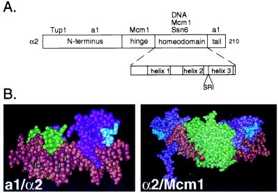
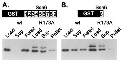
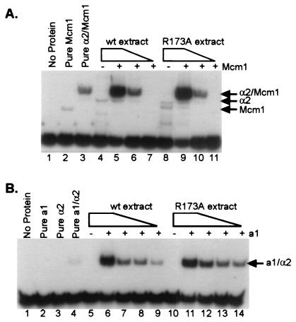
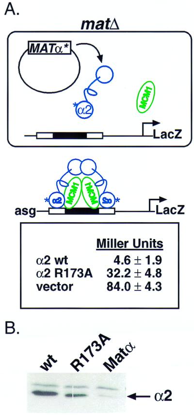
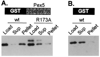
The tetratricopeptide repeat (TPR) is a 34-aa sequence motif, typically found in tandem clusters, that occurs in proteins of bacteria, archea, and eukaryotes. TPRs interact with other proteins, although few details on TPR-protein interactions are known. In this paper we show that a portion of a loop in the homeodomain of the DNA-binding protein alpha2 is required for its recognition by the TPRs of the corepressor Ssn6. The amino acid sequence of this loop is similar to the sequences recognized by the TPRs of an entirely different protein, Pex5, which directs peroxisomal import. We further show that alpha2 can be made to bind specifically in vitro to the TPRs of Pex5 and that a point mutation that disrupts the alpha2-Ssn6 interaction also disrupts the alpha2-Pex5 interaction. These results demonstrate that two different TPR proteins recognize their target by a similar mechanism, raising the possibility that other TPR-target interactions could occur through the same means.


Figures

Figure 1
Figure 1
α2 contains a surface exposed PTS1-like sequence in the homeodomain. (A) A schematic diagram of α2 illustrating its domain structure. Interacting proteins are placed above the domain where they are known to make contacts. The homeodomain is shown in more detail in the Inset. The PTS1-like sequence is placed below its position in the linear sequence of the homeodomain. (B) The PTS1-like sequence in the homeodomain of α2 is surface exposed. (Left) The structure of the α2 homeodomain (purple) is shown bound as a trimeric complex with the homeodomain of a1 (green) and DNA (brown) (21). (Right) The α2 homeodomain (purple) is shown bound to DNA (brown) and a dimer of Mcm1 (green) (20). In each of the structures the PTS1 sequence in the homeodomain of α2 is shown in blue.
Figure 2
Figure 2
An intact PTS1 sequence in the homeodomain of α2 is required for TPR binding. Wild-type or mutant (R173A) α2 extracts (Load) were incubated with resin containing immobilized GST-TPRs. After the incubation, the supernatant (Sup) contains unbound protein, and bound protein is pelleted with the resin and later eluted (Pellet). Equivalent volumes of sample were loaded in all lanes of a SDS/PAGE gel. Shown are Western blots of these gels probed with anti-α2 antibody. (A) This blot shows the result of incubating bacterial extracts containing either partially purified wild-type α2 or α2 R173A with resin containing immobilized GST:Ssn6 TPRs 1–9. (B) The resin contained immobilized GST:Ssn6 TPR6. The experiment of A was conducted at a final salt concentration of 175 mM, whereas the experiment in B was conducted at 150 mM final salt. The band of larger molecular weight present in the lanes containing α2 R173A is a covalent dimer of α2. The fraction of covalent dimer present in wild-type preparations is known to vary (39) and because the R173A protein behaves like the wild-type with respect to DNA binding and protein–protein interactions with a1 and Mcm1 (see below) we do not believe the presence of this dimer affects the interpretation of these experiments.
Figure 3
Figure 3
α2 R173A binds to DNA cooperatively and with wild-type (wt) affinity with its partners, Mcm1 and a1. (A) Gel mobility-shift assay using a Ste6 (a-specific gene) operator as probe. The gel mobility shifts shown here were produced by using decreasing amounts of either wild-type α2 or α2 R173A in combination with constant levels of purified Mcm11–96. Lane 1 contains free probe, and lane 2 contains added Mcm11–96 (2.3 × 10⁻⁹ M). All other lanes with added Mcm11–96 contain 4.6 × 10⁻¹⁰ M Mcm1 (lanes 3, 5–7, and 9–11). In addition to Mcm1, lane 3 contains purified α2 (1 × 10⁻⁸M). Lanes 5–7 and 9–11 contain 10-fold serial dilutions of either wild-type α2 or α2 R173A, respectively. The dilution series begins at 5 × 10⁻⁸ M and ends at 5 × 10⁻¹⁰ M, as indicated by the gradient above the lanes. Lanes 4 and 8 each contain 5 × 10⁻⁸ M α2 or α2 R173A, respectively, in the absence of any added Mcm11–96. (B) Gel mobility-shift assay using a haploid-specific gene operator as probe. The gel mobility shifts shown here were produced by using decreasing amounts of either wild-type α2 or α2 R173A in combination with constant levels of purified a1. Lane 1 contains free probe, lane 2 contains added purified a1 (8.4 × 10⁻⁸ M) in the absence of α2, and lane 3 contains purified α2 (1 × 10⁻⁹ M) in the absence of added a1. Lane 4 contains both purified a1 (8.4 × 10⁻⁸ M) and purified α2 (2 × 10⁻⁹ M). All other lanes with added a1 contain 8.4 × 10⁻⁸ M a1. Lanes 6–9 and 11–14 contain 2-fold serial dilutions of either wild-type α2 or α2 R173A, respectively. The dilution series begins at 1.6 × 10⁻⁹ M and concludes at 2.1 × 10⁻¹⁰ M, as indicated by the gradient above the lanes. Lanes 5 and 10 each contain 1.6 × 10⁻⁹ M wild-type α2 or α2 R173A, respectively, in the absence of added a1. Extract dilutions for each of the above gel mobility shifts were compared by Western blot to ensure that roughly equivalent levels of protein were used for each set of matched lanes (data not shown).
Figure 4
Figure 4
The PTS1 in the homeodomain of α2 is required for repression of a-specific genes. (A) (Upper) A schematic diagram illustrating the reporter gene system used in this experiment to quantitate repression. A matΔ strain contains a reporter gene construct integrated into the chromosome. In the reporter, upstream regulatory sequences control the activity of the LacZ gene. Here, these regulatory sequences contain an α2/Mcm1 operator. For repression to occur, α2 must bind its operator with Mcm1. In the absence of repression, the strain expresses high levels of β-galactosidase (vector). (Lower) Data from liquid β-galactosidase assays, effectively quantitating the amount of repression. In these experiments, we have conducted liquid β-galactosidase assays, in triplicate, on three unique transformants. Our data agrees with that previously published in ref. . (B) α2 and α2 R173A are expressed at comparable levels within the cells. Western blots of whole-cell extracts from the strains used in the liquid β-galactosidase assays above were probed with anti-α2 antibody. wt, wild type.
Figure 5
Figure 5
The PTS1-like sequence in α2 is required for the interaction with the TPRs of Pex5. The experiment was conducted as those in Fig. 2 with the exception that in A the resin contained immobilized GST:Pex5 TPRs 2–8, schematized by shading the TPRs. (B) This experiment serves as a binding control. In this experiment the resin contained only immobilized GST. The experiment of B was conducted at a final salt concentration of 175 mM, whereas the experiment in A was conducted at 150 mM final salt.

References

    1. Hirano T, Kinoshita N, Morikawa K, Yanagida M. Cell. 1990;60:319–328. - PubMed
    1. Sikorski R S, Boguski M S, Goebl M, Hieter P. Cell. 1990;60:307–317. - PubMed
    1. Lamb J R, Tugendreich S, Hieter P. Trends Biochem Sci. 1995;20:257–259. - PubMed
    1. Smith R L, Redd M J, Johnson A D. Genes Dev. 1995;9:2903–2910. - PubMed
    1. Terlecky S R, Nuttley W M, McCollum D, Sock E, Subramani S. EMBO J. 1995;14:3627–3634. - PMC - PubMed

Publication types

MeSH terms