. 2000 Apr;120(1):209-17.
doi: 10.1046/j.1365-2249.2000.01195.x.

Association of neutropenia in systemic lupus erythematosus (SLE) with anti-Ro and binding of an immunologically cross-reactive neutrophil membrane antigen

Affiliations

Association of neutropenia in systemic lupus erythematosus (SLE) with anti-Ro and binding of an immunologically cross-reactive neutrophil membrane antigen

B T Kurien et al. Clin Exp Immunol. 2000 Apr.

Abstract

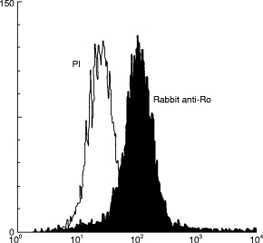
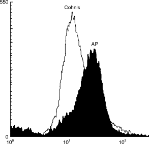
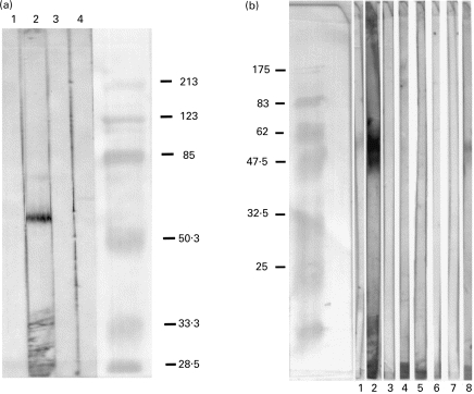
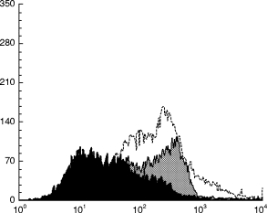
SLE is associated with the production of autoantibodies to self-constituents. In particular, certain ribonucleoprotein particles are targeted. Despite the multitude of autoantibodies produced and the remarkable concentrations of these antibodies in the sera of SLE patients, there have been little data that the autoantibodies found in SLE are involved in the pathogenesis of disease or its manifestations. The present work demonstrates that anti-Ro (or SSA) is associated with granulocytopenia, binds the surface of granulocytes and fixes complement to this membrane surface. Binding is a property of anti-Ro Fab fragments and can be inhibited by 60-kD Ro. However, the antigen bound on the surface of granulocytes is a 64 000 mol. wt protein that is a novel autoantigen in SLE. As suggested by inhibition studies, sequence identity between 60-kD Ro and eight tandem repeats in the 64-kD antigen may be responsible for the observed serologic cross-reactivity. These data imply that anti-Ro antibodies that also bind the 64-kD protein mediate neutropenia in patients with SLE.


Figures

Fig. 1
Fig. 1
Flow cytometry analysis of sera from normal persons as well as SLE patients with and without anti-Ro binding to human neutrophils. Human sera with equal amounts of protein based on A280 were added to the granulocytes and assayed for binding to the neutrophil surface as given in Patients and Methods. (a) Two anti-Ro SLE sera and two anti-Ro SLE sera binding to neutrophils. (b) A normal serum and four neutropenic anti-Ro sera analysed for binding to neutrophils.
Fig. 2
Fig. 2
Flow cytometry analysis of granulocyte binding by 60-kD Ro-immunized rabbit sera and pre-immune rabbit sera. FACS analysis of cells incubated with the pre-immune sera is shown by the open graph and fluorescence by cells incubated with 60-kD Ro-immunized rabbit sera is shown by the shaded graph.
Fig. 3
Fig. 3
Flow cytometry analysis of affinity-purified human anti-Ro (AP) binding to neutrophils. Cohn Fraction II (Cohn’s) at four times the concentration of the anti-Ro or purified anti-Ro was incubated with neutrophils and bound fluorescence was measured.
Fig. 4
Fig. 4
(a) Western immunoblot of granulocyte membrane prepared via nitrogen cavitation and probed with control or 60-kD Ro-immunized rabbit sera. Lane 1, Conjugate control; lane 2, 60-kD immunized rabbit; lane 3, rabbit pre-immune sera; lane 4, Freund’s adjuvant-immunized rabbit sera. (b) Western immunoblot of biotinylated granulocyte cell membrane. Granulocyte membrane-bound proteins were biotinylated and analysed on SDS–PAGE, followed by immunoblot. Lane 1, Pre-immune rabbit sera; lane 2, 60-kD Ro-immunized rabbit sera; lane 3, conjugate control; lane 4, normal human sera; lane 5, SLE sera with anti-Sm; lane 6, SLE sera without anti-Ro; lane 7, anti-P protein sera; lane 8, anti-Ro human sera.
Fig. 5
Fig. 5
Inhibition of anti-Ro binding intact, living neutrophils with 60-kD Ro MAP-230. Binding of anti-Ro to neutrophils is shown without inhibition (shaded grey), after incubation of the antibody with 10 μg of 60-kD Ro MAP-230 (shaded black), and after incubation with 10 μg of Ro-MAP-331 (unshaded), representing another sequence from 60-kD Ro. Sm-MAP-115 with residues 115–122 from Sm-B/B′ antigen served as another control and produced no inhibition (not shown).

References

    1. Nossent JC, Swaak AJG. Prevalence and significance of haematological abnormalities in patients with systemic lupus erythematosus. Quart J Med. 1991;80:605–12. - PubMed
    1. Goeckerman WH. Lupus erythematosus as a systemic disease. JAMA. 1923;80:542–7.
    1. Michael SR, Vural IL, Bassen FA, Schaefer L. The hematological aspects of disseminated (systemic) lupus erythematosus. Blood. 1951;6:1059–72. - PubMed
    1. Harvey AM, Shulman LE, Tumulty PA, Conley CL, Schoenrich EH. Systemic lupus erythematosus: review of the literature and clinical analysis of 138 cases. Medicine (Baltimore) 1954;33:291–437. - PubMed
    1. Starkebaum G, Arend WP. Neutrophil-binding immunoglobulin G in systemic lupus erythematosus. Evidence for the presence of both soluble immune complexes and immunoglobulin G antibodies to neutrophils. J Clin Invest. 1979;64:902–12. - PMC - PubMed

MeSH terms