. 2000 Apr 11;97(8):3884-9.
doi: 10.1073/pnas.97.8.3884.

Matrix metalloproteinase-2 is required for the switch to the angiogenic phenotype in a tumor model

Affiliations

Matrix metalloproteinase-2 is required for the switch to the angiogenic phenotype in a tumor model

J Fang et al. Proc Natl Acad Sci U S A. .

Abstract

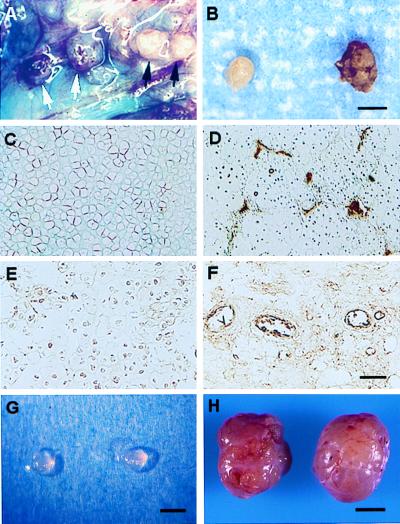
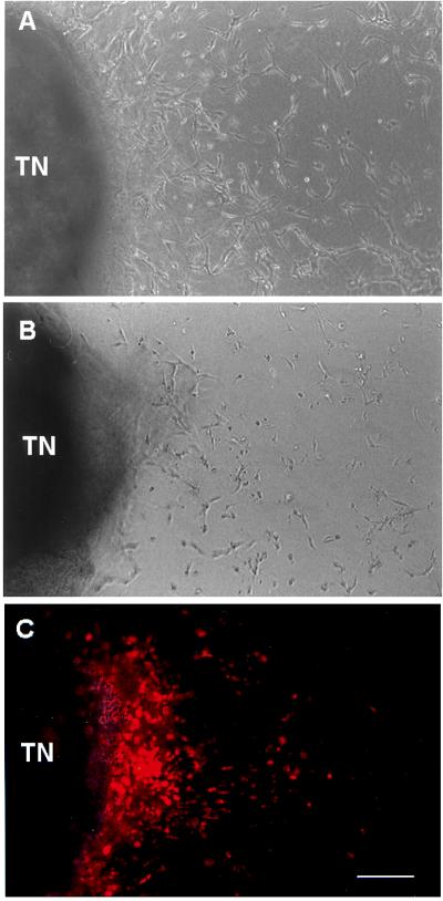
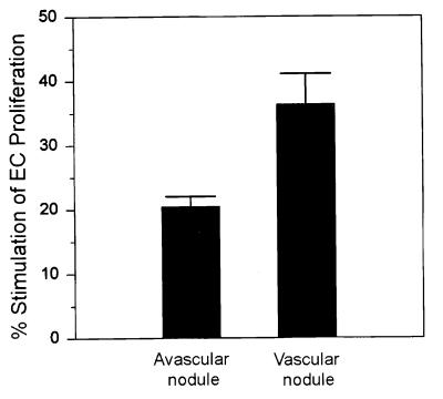
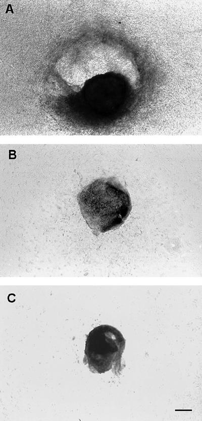
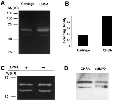
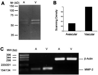
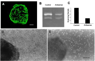
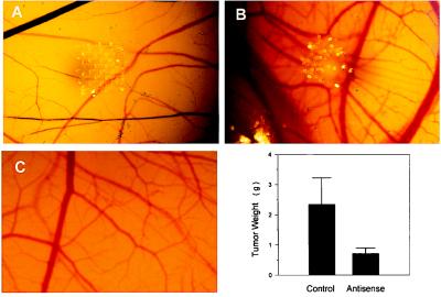
Among the earliest and most important stages during tumorigenesis is the activation of the angiogenic process, an event that is termed the "switch to the angiogenic phenotype." We have developed an in vivo system that can reliably recapitulate the stages in tumor development that represent this transition. Using this model, we have harvested and studied tumor nodules that can be distinguished from each other on the basis of their degree of vascularization. Angiogenic tumor nodules were characterized by the presence of capillary vessels as determined by factor VIII immunohistochemistry, and both angiogenic and proteolytic activities in vitro. In contrast, preangiogenic nodules were devoid of microvessels and showed little angiogenic or proteolytic activity in vitro. Addition of a specific metalloproteinase inhibitor resulted in the abrogation of both angiogenic and proteolytic activities of the angiogenic nodules in vitro. Comparative substrate gel electrophoresis detected the presence of a prominent matrix metalloproteinase (MMP-2) in the angiogenic nodules when compared with the preangiogenic ones. Suppression of MMP-2 activity by antisense oligonucleotides in the vascular nodules resulted in the loss of angiogenic potential both in vitro and in vivo in the chick chorioallantoic membrane assay. Moreover, this suppression of MMP-2 activity in angiogenic nodules inhibited tumor growth in vivo by approximately 70%. These results strongly implicate the activity of MMP-2 as a requirement for the switch to the angiogenic phenotype and validate this model as a reliable and reproducible tool by which to study other cellular and biochemical factors involved in the acquisition of the angiogenic phenotype.


Figures

Figure 1
Figure 1
Distinctive avascular and vascular phenotypes of chondrosarcoma tumor nodules. (A) Injection of tumor cell suspensions into air sacs in the sacral region of rats resulted in the formation of distinct tumor nodules, which could be visually distinguished from each other on the basis of their degree of vascularization i.e., vascular (white arrows) from avascular (black arrows). At day 10, approximately 50% of the tumor nodules were vascularized and by day 12, the majority of the nodules were vascularized. These results were consistently observed throughout more than 15 independent experiments. (B) Representative avascular and vascular nodules, respectively, are shown. Factor VIII immunostaining demonstrates the avascularity of representative control xiphoid cartilage sections (C) in which microvessels are not detected, in contrast to sections of chondrosarcoma in which a number of microvessels are easily seen (D). A lack of microvessels is observed in avascular chondrosarcoma nodules (E) when compared with vascular nodules in which positive microvessel staining is apparent (F). When avascular tumor nodules (G) were dissected from air sacs and implanted s.c. in rats, the majority of avascular nodules (11/13) became vascularized tumors (H). (Scale bars: A and B, 2 mm; C–F, 100 μm; G, 2 mm; H, 10 mm.)
Figure 2
Figure 2
In vitro angiogenesis assay of vascular and avascular chondrosarcoma tumor nodules. Vascular tumor nodules (TN), implanted in collagen gels throughout which capillary EC were dispersed, induced the concentration and alignment of capillary EC around the nodule (A) by day 3 in culture, in contrast to the lack of angiogenic response to avascular tumor nodules (B). When capillary EC as in A are labeled with fluoresceinated low density lipoprotein, the corona of EC around the vascular nodules is highlighted (C). To show the position of the vascular tumor nodule that is not fluorescent, the nodule in C is shown as a superimposed image taken with bright-field illumination. (Scale bar: A–C, 150 μm.)
Figure 3
Figure 3
Comparison of EC mitogenic activity between avascular and vascular tumor nodules. Protein was extracted from nodules, dialyzed against PBS, and added (10 μg/well) to each well of capillary EC. Stimulation of EC proliferation by nodule extracts was expressed in comparison to maximal stimulation (100%) by basic fibroblast growth factor (1 ng/ml, 0.4 ng/well), which resulted in a 4.2-fold increase in proliferation. All assays were done in duplicates. This result is representative of three independent experiments.
Figure 4
Figure 4
Comparison of proteolytic activity between vascular and avascular tumor nodules in vitro. By day 6 in culture, significant degradation of the collagen gel surrounding the vascular nodules was apparent (A), in contrast to gels surrounding the avascular nodules that remained intact (B). When 1,10-phenanthroline (5 μM) was added to the media overlaying the collagen gels in which vascular nodules were implanted, collagen gel degradation was completely suppressed (C). This result is representative of two independent assays (n = 7 for each treatment group). (Scale bar: AC, 400 μm.)
Figure 5
Figure 5
MMP-2 activity is increased in chondrosarcoma tissue in comparison to cartilage control. Zymography revealed predominant proteolytic bands migrating at molecular weights consistent with their identification as MMP-2 species. (A) Levels of this proteolytic activity were greater in chondrosarcoma (CHSA) than in the xiphoid cartilage controls. (B) Densitometric analysis documents an approximate 3-fold increase in intensity in chondrosarcoma as compared with the cartilage control. (C) Treatment of tumor extracts with 4-aminophenylmercuric acetate (APMA) showed no difference in the molecular mass of the enzyme species, suggesting that they are both present in their active forms. (D) Identification of enzyme activities as being those of MMP-2 was verified by immunoblot analysis using monospecific MMP-2 antibodies. Human recombinant MMP-2 (rMMP2) was included as a positive control.
Figure 6
Figure 6
MMP-2 activity is up-regulated in vascular tumor nodules. (A and B) Zymographic analysis revealed that the MMP-2 levels were three times greater in vascular tumor nodules than in avascular nodules. RT-PCR analysis (C) demonstrated a significantly higher MMP-2 expression level in vascular nodules (V) when compared with avascular nodules (A). β-actin served as an internal control.
Figure 7
Figure 7
Inhibition of MMP-2 expression and angiogenesis by antisense oligonucleotides in vitro. (A) Uptake of oligonucleotides by tumor cells in vitro was demonstrated by cellular localization of FITC-labeled oligonucleotides in vascular tumor nodules. (B and C) Treatment of vascular nodules with MMP-2 antisense oligonucleotides resulted in a 70% reduction in MMP-2 activity in the conditioned media in comparison to that of nodules treated with control oligonucleotides. Capillary EC responded to tumor nodules in the presence of control oligonucleotides (D), but did not in the presence of 10 μM MMP-2 antisense oligonucleotides (E). (Scale bars: A, 250 μm; D and E, 200 μm.)
Figure 8
Figure 8
Suppression of angiogenesis and tumor growth by MMP-2 antisense in vivo. Vascular tumor nodules preincubated with 10 μM oligonucleotides inhibited the normal embryonic vasculature of the chick CAM (5/5) (A), in contrast to the control nodules that were pretreated with random oligonucleotides and that stimulated capillary growth (4/5) (B), and compared with untreated CAM control (C). Vascular tumor nodules that were preincubated with 10 μM MMP-2 antisense for 3 days and were implanted into animals (n = 6) resulted in 69% suppression of tumor growth in comparison to control nodules treated with random oligonucleotides (P < 0.05).

References

    1. Hanahan D, Folkman J. Cell. 1996;86:353–364. - PubMed
    1. Nagase H, Woessner J F., Jr J Biol Chem. 1999;274:21491–21494. - PubMed
    1. Cockett M I, Murphy G, Birch M L, O'Connell J P, Crabbe T, Millican A T, Hart I R, Docherty A J. Biochem Soc Symp. 1998;63:295–313. - PubMed
    1. Kahari V M, Saarialho-Kere U. Ann Med. 1999;31:34–45. - PubMed
    1. Kleiner D E, Stetler-Stevenson W G. Cancer Chemother Pharmacol. 1999;43,Suppl.:42–51. - PubMed

Publication types

LinkOut - more resources