. 2000 Apr 11;97(8):4017-22.
doi: 10.1073/pnas.97.8.4017.

The polycystic kidney disease protein PKD2 interacts with Hax-1, a protein associated with the actin cytoskeleton

Affiliations

The polycystic kidney disease protein PKD2 interacts with Hax-1, a protein associated with the actin cytoskeleton

A R Gallagher et al. Proc Natl Acad Sci U S A. .

Abstract

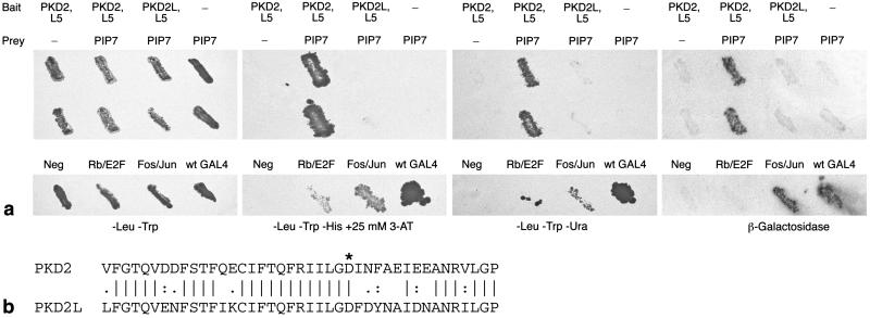
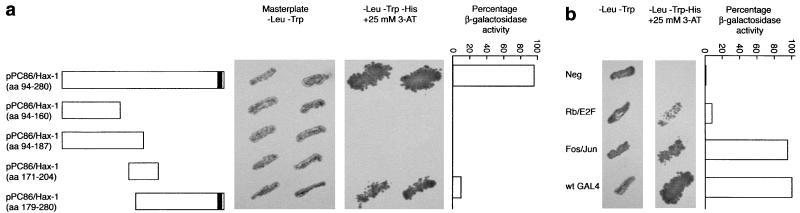
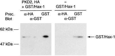
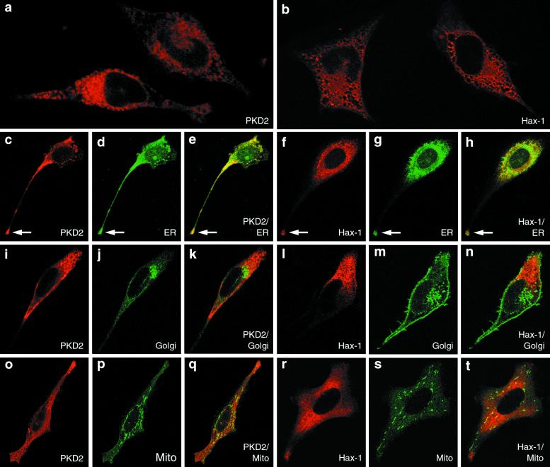
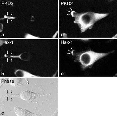
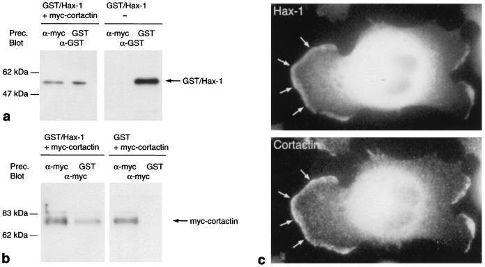
Despite the recent positional cloning of the PKD1 and PKD2 genes, which are mutated in the great majority of patients with autosomal-dominant polycystic kidney disease (PKD), the pathogenic mechanism for cyst formation is still unclear. The finding, that the PKD1 and PKD2 proteins interact with each other through their COOH termini, suggests that both proteins are part of the same protein complex or signal transduction pathway. Using a yeast two-hybrid screen with the PKD2 protein, we isolated the PKD2-interacting protein Hax-1. The specificity of the interaction was demonstrated by the fact that PKD2L, a protein closely related to PKD2, failed to interact with Hax-1. Immunofluorescence experiments showed that in most cells PKD2 and Hax-1 colocalized in the cell body, but in some cells PKD2 and Hax-1 also were sorted into cellular processes and lamellipodia. Furthermore we demonstrated an association between Hax-1 and the F-actin-binding protein cortactin, which suggests a link between PKD2 and the actin cytoskeleton. We speculate that PKD2 is involved in the formation of cell-matrix contacts, which are dysfunctional without a wild-type PKD2 protein, thus leading to cystic enlargement of tubular structures in the kidney, liver, and pancreas.


Figures

Figure 1
Figure 1
Two-hybrid assays for the interaction between PKD2 and Hax-1. (a) Using three different assays it can be seen that Hax-1 interacts strongly and specifically with PKD2. Both in an assay for histidine and uracil prototrophy and in a β-galactosidase assay the region between the fifth and sixth transmembrane domain of PKD2 (loop 5; PKD2, L5) scores positive when expressed together with a truncated Hax-1 protein (PIP7). Loop 5 of the closely related PKD2L protein, however, fails to interact with Hax-1 (PKD2L, L5). The bottom panels show the negative and positive controls either containing no bait and no prey (Neg), a bait/prey combination of the retinoblastoma protein (Rb) and E2F (Rb/E2F), a bait/prey combination of Fos and Jun (Fos/Jun), and the wild-type GAL4 protein (wt GAL4). The master plates without additional selection are shown on the left. (b) Sequence comparison of loop 5 in human PKD2 and PKD2L. The negatively charged (aspartic acid) residue, which is considered to be important for cation selectivity in Ca2+ channels, is indicated by *.
Figure 2
Figure 2
The COOH terminus of Hax-1 mediates interaction with PKD2. The yeast two-hybrid system was used to determine the region in Hax-1 necessary for the interaction with loop 5 of PKD2. An interaction was assayed by prototrophy for histidine in the presence of 25 mM 3-aminotriazole and by a liquid β-galactosidase assay (the β-galactosidase activity achieved with the wild-type GAL4 protein was set at 100%). Although the last 102 aa of Hax-1 are sufficient for the association with PKD2, this interaction is not as strong as the one seen with the longer Hax-1 fragment. Therefore additional amino acids are probably necessary as part of the domain interacting with PKD2 or to maintain the appropriate conformation of Hax-1 necessary to interact with PKD2. The putative transmembrane domain of Hax-1 is depicted as a vertical bar at the COOH terminus.
Figure 3
Figure 3
Coprecipitation of PKD2 and Hax-1 in mammalian cells. Protein extracts from stably transfected HeLa cells inducibly expressing a HA epitope-tagged PKD2 protein and constitutively expressing a GST/Hax-1 fusion protein were incubated with the anti-HA epitope antibody 12CA5 (Prec.). Immunoprecipitated proteins were analyzed by Western blot with an anti-GST antibody (Blot). The GST/Hax-1 fusion protein is precipitated only in extracts from those cells where the expression of PKD2 is turned on (Left), but not in extracts from cells where the expression of PKD2 is turned off (Right). As a positive control the GST/Hax-1 fusion protein was precipitated with glutathione-agarose beads (GST).
Figure 4
Figure 4
PKD2 and Hax-1 are located in the endoplasmic reticulum. HeLa cells either inducibly expressing a HA epitope-tagged PKD2 protein or constitutively expressing a GST/Hax-1 fusion protein showed a reticular cytoplasmic distribution of both proteins when stained with the anti-HA epitope antibody (a) or with an anti-GST antibody (b). Double-staining with markers for the endoplasmic reticulum (anti-Sec61 β antibody, ch), the Golgi apparatus (anti-Golgi 58K protein antibody, ik; FITC-conjugated wheat germ agglutinin, ln) and mitochondria (anti-Mn-superoxide dismutase antibody, ot) demonstrates that PKD2 and Hax-1 are located in the endoplasmic reticulum (ch), but not in the Golgi apparatus (in) nor in the mitochondria (ot). The arrows in ch point to the tips of long cell processes where PKD2 and Hax-1 were detected. Note that with the FITC-conjugated wheat germ agglutinin the outline of the cell also is stained because of the presence of N-glycosylated plasma membrane proteins (m and n).
Figure 5
Figure 5
Colocalization of PKD2 and Hax-1 in mammalian cells. Stably transfected HeLa cells inducibly expressing PKD2 and constitutively expressing a GST/Hax-1 fusion protein were double-stained with the polyclonal anti-PKD2 antibody (a and d) and a monoclonal anti-GST antibody (b and e). In most cells PKD2 and Hax-1 colocalize in the cytoplasm, but in those cells spreading out over the dish a striking redistribution into the tips of the cell processes (arrows) could be seen (ac). A phase contrast view is included to better demonstrate the shape of the cell (c). In very few cells both proteins were sorted into lamellipodia (arrows in d and e). PKD2 and Hax-1 only partially colocalize in the body of the cell shown in d and e, suggesting a dynamic interaction between PKD2 and Hax-1.
Figure 6
Figure 6
Hax-1 is associated with cortactin. (a and b) COS-7 cells were transiently transfected with various combinations of expression plasmids coding for myc-tagged cortactin, a GST/Hax-1 fusion protein and GST only. Protein extracts from transfected cells were used for immunoprecipitation with an anti-myc-epitope antibody and for GST pull-down assays (Prec.). The precipitates then were analyzed by Western blot with an anti-GST (a) and anti-myc-epitope antibody (b) (Blot). In either case cortactin and Hax-1 are coprecipitated, demonstrating an association between the two proteins. (c) Stably transfected HeLa cells constitutively expressing a GST/Hax-1 fusion protein were double-stained with a mouse mAb against GST (Upper) and a rabbit polyclonal antibody against cortactin (Lower). It can be easily seen that the two proteins colocalize in lamellipodia (arrows).

References

    1. Gabow P A. N Engl J Med. 1993;329:332–342. - PubMed
    1. The European Polycystic Kidney Disease Consortium. Cell. 1994;77:881–894. - PubMed
    1. Mochizuki T, Wu G, Hayashi T, Xenophontos S L, Veldhuisen B, Saris J J, Reynolds D M, Cai Y, Gabow P A, Pierides A, et al. Science. 1996;272:1339–1342. - PubMed
    1. Hughes J, Ward C J, Peral B, Aspinwall R, Clark K, San Millán J L, Gamble V, Harris P C. Nat Genet. 1995;10:151–159. - PubMed
    1. The International Polycystic Kidney Disease Consortium. Cell. 1995;81:289–298. - PubMed

Publication types

MeSH terms