. 2000 Apr 11;97(8):4297-302.
doi: 10.1073/pnas.97.8.4297.

Genetic evidence that the bacteriophage phi X174 lysis protein inhibits cell wall synthesis

Affiliations

Genetic evidence that the bacteriophage phi X174 lysis protein inhibits cell wall synthesis

T G Bernhardt et al. Proc Natl Acad Sci U S A. .

Abstract

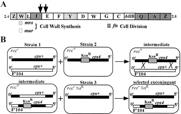
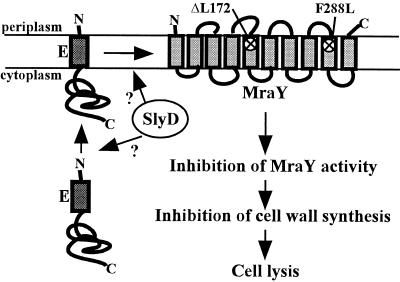
Protein E, a 91-residue membrane protein of phiX174, causes lysis of the host in a growth-dependent manner reminiscent of cell wall antibiotics, suggesting E acts by inhibiting peptidoglycan synthesis. In a search for the cellular target of E, we previously have isolated recessive mutations in the host gene slyD (sensitivity to lysis) that block the lytic effects of E. The role of slyD, which encodes a FK506 binding protein-type peptidyl-prolyl cis-trans isomerase, is not fully understood. However, E mutants referred to as Epos (plates on slyD) lack a slyD requirement, indicating that slyD is not crucial for lysis. To identify the gene encoding the cellular target, we selected for survivors of Epos. In this study, we describe the isolation of dominant mutations in the essential host gene mraY that result in a general lysis-defective phenotype. mraY encodes translocase I, which catalyzes the formation of the first lipid-linked intermediate in cell wall biosynthesis. The isolation of these lysis-defective mutants supports a model in which translocase I is the cellular target of E and that inhibition of cell wall synthesis is the mechanism of lysis.


Figures

Figure 1
Figure 1
(A) The mra locus. Shown is a schematic of the mra locus of E. coli present at 2 min. The positions of the mini-Tncam insertions that reduce the ability of F′104eps4 to confer the dominant Eps phenotype are indicated by the arrows. (B) Triparental mating strategy to generate merodiploids. Shown is a schematic of the triparental mating strategy used to generate merodiploids with the eps+ allele on the chromosome and the eps4 allele on F′104. The thick black line represents the 0- to 5-min region of the chromosome. The open line represents the 0- to 5-min region contained on F′104. Relevant alleles present in the 0- to 5-min regions are indicated above the lines. All other relevant genotypic features are indicated in the top left corner of the cartoon cells. The shaded box represents the transposon Tn10kan present at 2 min. The crossed lines represent homologous recombination between the F′ and chromosome. The same strategy was used to generate homozygous eps+ merodiploids by using a strain 2 with eps+ linked to the KanR transposon marker.
Figure 2
Figure 2
The mraY4 allele causes a general lysis defect. (A) Lysis profiles from pEmycZ induction in different genetic backgrounds. The allelic state of slyD and mraY in each strain is as follows: slyD+ mraY+ (RY7425) (○), slyD1 mraY+ (RY7426) (□), slyD+ mraY4 (RY7427) (▵), and slyD1 mraY4 (RY7428) (+). The arrow indicates the time of IPTG addition to induce lysis gene expression. (B) The strains are the same as indicated in A except they contain the plasmid pEposmycZ. (C) Same as in A except strains contain the plasmid pE35GFP. (D) Lysis profiles resulting from increased mraY expression. Emyc expression was induced from CAG12095 pBAD30 pEmycZK (○), pMY30 pEmycZK (□), and pMY304 pEmycZK (▵) at time 0. All cultures were grown in the presence of 0.2% arabinose for constitutive expression of the mraY alleles cloned in the pBAD30 vector.
Figure 3
Figure 3
Proposed model for the mechanism of E-mediated cell lysis. The E protein is synthesized in the cytoplasm and integrated into the inner membrane. The cytoplasmic SlyD PPIase is required for E protein accumulation, either before or after E reaches the membrane. We propose that as E accumulates in the membrane it interacts with and inhibits MraY. Inhibition of MraY activity would result in the inhibition of cell wall synthesis and cell lysis. The predicted topology of MraY in the membrane is drawn with the positions of the mutations affecting lysis indicated with an x. The topology of the E protein is drawn according to the observation that the C terminus is located in the cytoplasm (R.Y., unpublished data), although it is not known whether the N terminus of E actually traverses the membrane.

References

    1. Young R. Microbiol Rev. 1992;56:430–481. - PMC - PubMed
    1. Sanger F, Air G M, Barrell B G, Brown N L, Coulson A R, Fiddes J C, Hutchison C A, III, Slocombe P M, Smith M. Nature (London) 1977;265:687–695. - PubMed
    1. Young K D, Young R. J Virol. 1982;44:993–1002. - PMC - PubMed
    1. Henrich B, Lubitz W, Plapp R. Mol Gen Genet. 1982;185:493–497. - PubMed
    1. Burman L G, Raichler J, Park J T. J Bacteriol. 1983;155:983–988. - PMC - PubMed

Publication types

LinkOut - more resources