. 2000 Mar 30;404(6777):510-4.
doi: 10.1038/35006670.

DNA repair protein Ku80 suppresses chromosomal aberrations and malignant transformation

Affiliations

DNA repair protein Ku80 suppresses chromosomal aberrations and malignant transformation

M J Difilippantonio et al. Nature. .

Abstract

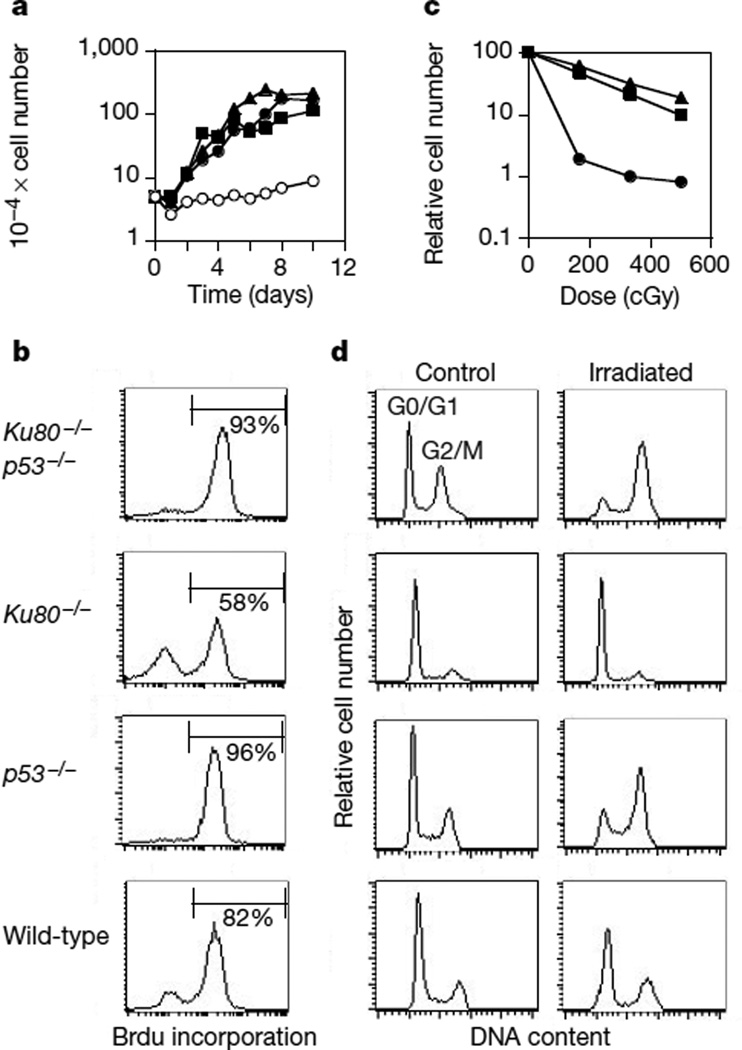
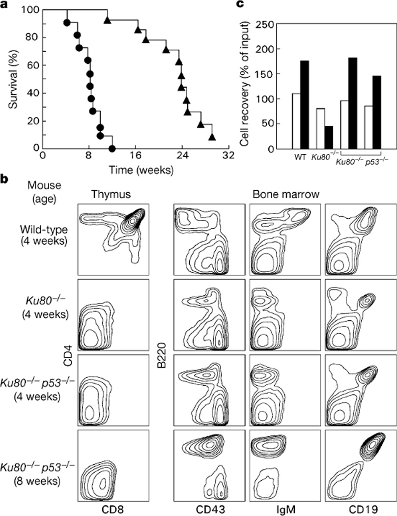
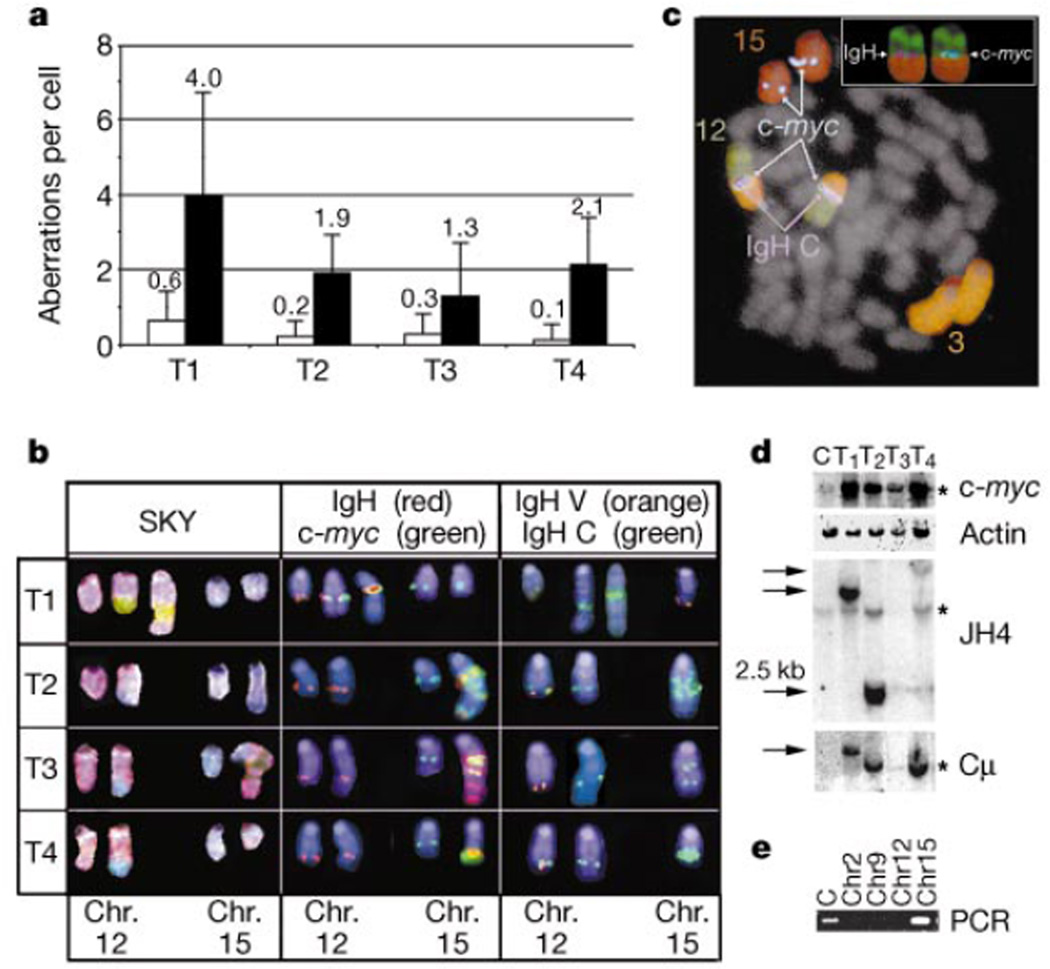
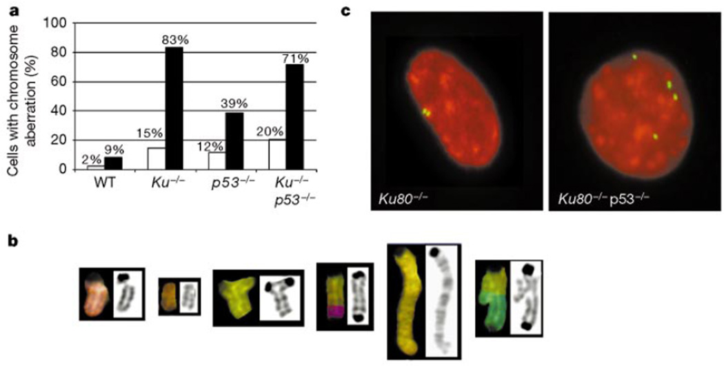
Cancer susceptibility genes have been classified into two groups: gatekeepers and caretakers. Gatekeepers are genes that control cell proliferation and death, whereas caretakers are DNA repair genes whose inactivation leads to genetic instability. Abrogation of both caretaker and gatekeeper function markedly increases cancer susceptibility. Although the importance of Ku80 in DNA double-strand break repair is well established, neither Ku80 nor other components of the non-homologous end-joining pathway are known to have a caretaker role in maintaining genomic stability. Here we show that mouse cells deficient for Ku80 display a marked increase in chromosomal aberrations, including breakage, translocations and aneuploidy. Despite the observed chromosome instabilities, Ku80-/- mice have only a slightly earlier onset of cancer. Loss of p53 synergizes with Ku80 to promote tumorigenesis such that all Ku80-/- p53-/- mice succumb to disseminated pro-B-cell lymphoma before three months of age. Tumours result from a specific set of chromosomal translocations and gene amplifications involving IgH and c-Myc, reminiscent of Burkitt's lymphoma. We conclude that Ku80 is a caretaker gene that maintains the integrity of the genome by a mechanism involving the suppression of chromosomal rearrangements.


Figures

Figure 1
Figure 1
Growth characteristics of untreated and irradiated mouse embryo fibroblasts. a, Growth kinetics of Ku80−/− (open circles), p53−/− (filled triangles), Ku80−/−p53−/− (filled circles) and wild-type (filled squares) MEFs. b, Incorporation of BrdU in MEF cultures after 16 h of continuous labelling. c, Radiation sensitivity of Ku80−/−p53−/− (circles), p53−/− (triangles) and wild-type (squares) MEFs, plotted as the fraction of surviving cells relative to unirradiated samples of the same genotype. Ku80−/− MEFs exhibited a sevenfold decrease in cell count independent of the dose (83–600 cGy) owing to the premature senescence of unirradiated cultures, and the permanent arrest of the dividing population in response to γ-irradiation. d, DNA content histogram measured in untreated samples (control) and in cultures 24 h after treatment with 10 Gy of γ-radiation (Irradiated).
Figure 2
Figure 2
Analysis of lifespan and lymphocyte development. a, Kaplan–Meier analysis comparing mortality of Ku80+/+p53−/− (triangles) and Ku80−/−p53−/− (circles) mice as a function of time. The survival of Ku80−/−p53−/− mice is statistically different from that of the Ku80+/+p53−/− group; P = 0.0001. b, Flow cytometric analysis of thymocyte (left panel) and bone-marrow (right panels) suspensions obtained from wild-type, Ku80−/− and Ku80−/−p53−/− mice. An 8-week-old Ku80−/−p53−/− mouse, which had a B-cell lymphoma, is compared with a 4-week-old non-tumour-bearing Ku80−/−p53−/− mouse, and with 4-week-old Ku80−/− and wild-type littermates. Data are representative of 10 wild-type, 6 Ku80−/−, 5 non-tumour-bearing Ku80−/−p53−/− mice 2–4 weeks old, and 12 Ku80−/−p53−/− animals that had tumours. Average thymus cellularity was 5 × 10⁵ in Ku80−/− mice, 106 in non-tumour-bearing Ku80−/−p53−/− mice and 2 × 10⁸ in wild-type controls. Average number of B220+CD19+ cells in bone marrow was 4.6 × 10⁵ in Ku80−/− mice, 9.6 × 10⁵ in non-tumour-bearing Ku80−/−p53−/− mice and 2.8 × 10⁶ for wild-type controls. The immuno-phenotypes of Ku80+/+p53−/− and wild-type mice were similar (not shown). c, Survival of wild-type (WT) B220+CD43+IgM pro-B cells, and Ku80−/−p53−/− and Ku80−/− CD19+ B cells after 1 d (open columns) and 3 d (filled columns) in culture, plotted relative to input cell number at day 0. Results from two independent Ku80−/−p53−/− mice are plotted.
Figure 3
Figure 3
c-myc and IgH-associated rearrangements and amplifications in Ku80−/−p53−/− tumours. a, Average number of translocations (other than those juxtaposing chromosomes 12 and 15), unshaded, or gains and loses of chromosomes, shaded, in Ku80−/−p53−/− tumours (T1–T4). b, SKY analysis revealed translocations involving chromosomes (Chr.) 12 (pink) and 15 (blue) in Ku80−/−p53−/− tumours 2–4 (T2–T4) and 12 and 3 (green) in T1. FISH with probes for IgH Cγ1−α (red) and c-myc (green) reveal co-localization and co-amplification of these genes as well as signals on the normal chromosomes. Analysis with probes for IgH variable (orange) and constant (green) regions show co-localization on the normal chromosome 12. The absence of the variable region on the derivative 12 shows that this translocation disrupts the IgH locus. Chromosomes in the middle and right panels were identified by hybridization with painting probes (as in c) and are counterstained with 4,6-diamidino-2-phenylindole (DAPI; blue). c, Five-colour FISH analysis of a metaphase from tumour T1. The inset shows the localization of individual c-myc and IgH signals at the breakpoint between chromosomes 12 and 3. d, Southern blot analysis showing germline bands (stars) as well as rearrangments (arrows) and/or amplifications of c-myc and IgH. e, PCR amplification of tumour 2 breakpoint indicates that the JH1 fusion partner originated from chromosome 15. Abbreviations: C: Liver; T1–T4: Ku80−/−p53−/− tumors 1–4.
Figure 4
Figure 4
Chromosome aberrations in Ku80−/− and Ku80−/−p53−/− MEFs. a, Percentage of cells with DNA breaks or translocations, shaded (n = 35), and polyploidy, unshaded (n = 400), observed in passage 1 MEFs. WT, wild type. b, Typical chromosome aberrations found in Ku80−/− MEFs. From left to right, chromatid break, acentric fragment, tri-radial, dicentric, duplication and an asymmetric exchange. c, γ-Tubulin staining for centrosomes (green) in Ku80−/− (left) and Ku80−/−p53−/− (right) MEFs counterstained with propidium iodide (red).

Comment in

  • New guardians of the genome.
    Roth DB, Gellert M. Roth DB, et al. Nature. 2000 Apr 20;404(6780):823-5. doi: 10.1038/35009180. Nature. 2000. PMID: 10786775 No abstract available.

Similar articles

Cited by

References

    1. Kinzler KW, Vogelstein B. Gatekeepers and caretakers. Nature. 1997;386:761–763. - PubMed
    1. Vogel H, Lim DS, Karsenty G, Finegold M, Hasty P. Deletion of Ku86 causes early onset of senescence in mice. Proc. Natl Acad. Sci. USA. 1999;96:10770–10775. - PMC - PubMed
    1. Nussenzweig A, et al. Requirement for Ku80 in growth and immunoglobulin V(D)J recombination. Nature. 1996;382:551–555. - PubMed
    1. Zhu C, Bogue MA, Lim DS, Hasty P, Roth DB. Ku86-deficient mice exhibit severe combined immunodeficiency and defective processing of V(D)J recombination intermediates. Cell. 1996;86:379–389. - PubMed
    1. Ouyang H, et al. Ku70 is required for DNA repair but not for T cell antigen receptor gene recombination in vivo. J. Exp. Med. 1997;186:921–929. - PMC - PubMed

Publication types

MeSH terms