. 2000 May;182(9):2445-52.
doi: 10.1128/JB.182.9.2445-2452.2000.

Efficient targeted mutagenesis in Borrelia burgdorferi

Affiliations

Efficient targeted mutagenesis in Borrelia burgdorferi

J L Bono et al. J Bacteriol. 2000 May.

Abstract

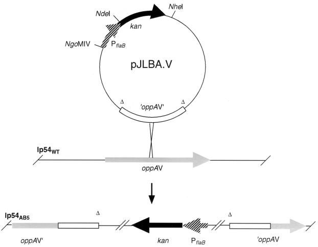
Genetic studies in Borrelia burgdorferi have been hindered by the lack of a nonborrelial selectable marker. Currently, the only selectable marker is gyrB(r), a mutated form of the chromosomal gyrB gene that encodes the B subunit of DNA gyrase and confers resistance to the antibiotic coumermycin A(1). The utility of the coumermycin-resistant gyrB(r) gene for targeted gene disruption is limited by a high frequency of recombination with the endogenous gyrB gene. A kanamycin resistance gene (kan) was introduced into B. burgdorferi, and its use as a selectable marker was explored in an effort to improve the genetic manipulation of this pathogen. B. burgdorferi transformants with the kan gene expressed from its native promoter were susceptible to kanamycin. In striking contrast, transformants with the kan gene expressed from either the B. burgdorferi flaB or flgB promoter were resistant to high levels of kanamycin. The kanamycin resistance marker allows efficient direct selection of mutants in B. burgdorferi and hence is a significant improvement in the ability to construct isogenic mutant strains in this pathogen.


Figures

FIG. 1
FIG. 1
Construction of pJLB500A and pJLB500G plasmids. (A) PCR fragments of B. burgdorferi promoters and the kan gene were cloned into pCR2.1. (B) The kan gene was excised and ligated into plasmids containing B. burgdorferi promoters. The resulting plasmids contained PflaB-kan or PflgB-kan fusions (pTAkanA and pTAkanG, respectively). (C) pTAkanAn and pTAkanGn plasmids were constructed by amplifying PflaB-kan and PflgB-kan from pTAkanA and pTAkanG, respectively, with primers that contained NgoMIV and NheI restriction sites. The PCR product amplified with oligonucleotides 10 and 11 from pBLS500 was cloned into pCR2.1 (pTA500). (D) PflaB-kan and pTA500 or PflgB-kan and pTA500 were digested, purified, and ligated to create pJLB500A or pJLB500G, respectively. The shaded box represents either the B. burgdorferi flaB or flgB promoter region. The black box and open box represent the kan and gyrBr genes, respectively. The thick line represents pOK12 sequence. Plasmids are not drawn to scale.
FIG. 2
FIG. 2
Sequence of the promoter regions from flaB and flgB of B. burgdorferi and the kan gene of Tn903. The NgoMIV site was added to the 5′ end of the flaB and flgB promoters for cloning. The underlined sequence denotes the NdeI fusion site between the promoters and the kan gene. The nucleotides shown in boldface within the underlined sequence differ from the wild-type sequence. The transcription initiation sites are indicated by arrows, and the proposed −10 and −35 sequences of the promoters are overlined (8, 9, 11, 14, 15). The first five amino acids of the kan product are shown below their corresponding codons.
FIG. 3
FIG. 3
Southern blot analysis of DNA from various B. burgdorferi clones. Total genomic DNA from clones B31-A (WT), AB1, KA1, and KG1 digested with XbaI and hybridized with either a gyrB (A) or a kan (B) probe (strain source of DNA indicated at the top of each lane). The size standards are indicated on the left.
FIG. 4
FIG. 4
Northern blot analysis of kan transcripts from B. burgdorferi clones. A blot containing total RNA from B. burgdorferi clones B31-A (WT), AB1, KA1, and KG1 was hybridized with a radioactive kan probe and exposed for 8 h at −80°C (A). After decay, these blots were hybridized with an internal flagellin probe to assure equivalent loading of RNA in each lane (B). An RNA size marker (in kilobases) is indicated to the left of each panel.
FIG. 5
FIG. 5
Formation of B. burgdorferi clone AB5. The top of the figure shows the plasmid pJLBA.V and integration at the oppAV locus in the 54-kb linear plasmid (lp54). The lower part of the figure represents the oppAV locus of the B. burgdorferi clone AB5 with an integrated copy of pJLBA.V.

Similar articles

Cited by

References

    1. Barbour A G. Isolation and cultivation of Lyme disease spirochetes. Yale J Biol Med. 1984;57:521–525. - PMC - PubMed
    1. Barbour A G, Hayes S F, Heiland R A, Schrumpf M E, Tessier S L. A Borrelia-specific monoclonal antibody binds to a flagellar epitope. Infect Immun. 1986;52:549–554. - PMC - PubMed
    1. Bono J L, Tilly K, Stevenson B, Hogan D, Rosa P. Oligopeptide permease in Borrelia burgdorferi: putative peptide-binding components encoded by both chromosomal and plasmid loci. Microbiology. 1998;144:1033–1044. - PubMed
    1. Burgdorfer W, Barbour A G, Hayes S F, Benach J L, Grunwaldt E, Davis J P. Lyme disease—a tick-borne spirochetosis? Science. 1982;216:1317–1319. - PubMed
    1. Carroll J A, Garon C F, Schwan T G. Effects of environmental pH on membrane proteins in Borrelia burgdorferi. Infect Immun. 1999;67:3181–3187. - PMC - PubMed

MeSH terms

LinkOut - more resources