. 2000 May;182(9):2461-7.
doi: 10.1128/JB.182.9.2461-2467.2000.

Dimers of pi protein bind the A+T-rich region of the R6K gamma origin near the leading-strand synthesis start sites: regulatory implications

Affiliations

Dimers of pi protein bind the A+T-rich region of the R6K gamma origin near the leading-strand synthesis start sites: regulatory implications

R Krüger et al. J Bacteriol. 2000 May.

Abstract

The replication of gamma origin, a minimal replicon derived from plasmid R6K, is controlled by the Rep protein pi. At low intracellular concentrations, pi activates the gamma origin, while it inhibits replication at elevated concentrations. Additionally, pi acts as a transcription factor (auto)repressing its own synthesis. These varied regulatory functions depend on pi binding to reiterated DNA sequences bearing a TGAGNG motif. However, pi also binds to a "non-iteron" site (i.e., not TGAGNG) that resides in the A+T-rich region adjacent to the iterons. This positioning places the non-iteron site near the start sites for leading-strand synthesis that also occur in the A+T-rich region of gamma origin. We have hypothesized that origin activation (at low pi levels) would require the binding of pi monomers to iterons, while the binding of pi dimers to the non-iteron site (at high pi levels) would be required to inhibit priming. Although monomers as well as dimers can bind to an iteron, we demonstrate that only dimers bind to the non-iteron site. Two additional pieces of data support the hypothesis of negative replication control by pi binding to the non-iteron site. First, pi binds to the non-iteron site about eight times less well than it binds to a single iteron. Second, hyperactive variants of pi protein (called copy-up) either do not bind to the non-iteron site or bind to it less well than wild-type pi. We propose a replication control mechanism whereby pi would directly inhibit primer formation.


Figures

FIG. 1
FIG. 1
Selected elements of γ ori and known or proposed functions of π. The seven 22-bp iterons (π binding sites) are indicated by black arrows. The non-iteron π binding and IHF binding sites (ihf1) are indicated in the A+T-rich segment adjacent to iterons. SSLSS are indicated by vertical arrows; the direction of replication is indicated by a horizontal gray arrow. pir is the structural gene for π protein. A pair of inverted arrows in the pir operator indicates the inverted repeats. The inhibition of replication and/or repression of pir gene transcription is indicated by a minus sign. The activation of replication is indicated by a plus sign.
FIG. 2
FIG. 2
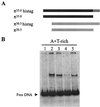
Binding of π protein variants to the non-iteron site. (A) Diagram of proteins examined by EMSA. (B) Binding reactions were assembled as described in Materials and Methods with a non-iteron DNA probe obtained from plasmid pTS5. The lanes represent non-iteron DNA probe without protein (lane 1) or with π35.0·His6 (lane 2), π35.0 (lane 3), π30.5·His6 (lane 4), and π30.5 (lane 5).
FIG. 3
FIG. 3
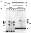
Oligomerization assay of π bound to iteron and non-iteron sites. (A) Diagram of proteins examined by EMSA for their oligomerization state in complexes with the iteron-containing probe (left panel) and the non-iteron probe (right panel). π35.0·His6 homodimers, π30.5 homodimers, and π35.0·His630.5 heterodimers were obtained as described in Materials and Methods. (B) The lanes represent probes without protein (lanes 1 and 5) or with π35.0·His6 (lanes 2 and 7); π30.5 (lanes 3 and 8); or a mixture of π35.0·His6 and π30.5 eluted from Ni2+-NTA resin (lanes 4 and 9), resulting in π35.0·His6 homodimers, π35.0·His630.5 heterodimers, and π35.0·His6 monomers. IHF alone (lane 6) or in combination with the π variants was added to samples containing the non-iteron probe (lanes 7 to 9).
FIG. 4
FIG. 4
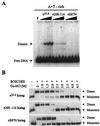
Assay for the number of π subunits sufficient for binding to the non-iteron site. (A) Diagram of proteins examined by EMSA for their binding to the non-iteron probe. Homodimers of π35.0·His6 and ΔC164π as well as π35.0·His6 ΔC164π heterodimers were obtained as described in Materials and Methods. (B) The lanes represent DNA probe without protein (lane 1) or with π35.0·His6 (lane 2); a mixture of π35.0·His6 and ΔC164π resulting in π35.0·His6-ΔC164π heterodimers, π35.0·His6 homodimers, and π35.0·His6 monomers (lane 3); or ΔC164π homodimers (lane 4).
FIG. 5
FIG. 5
Comparison of π35.0·His6 binding to the iteron versus non-iteron sites. π35.0·His6 was used with a direct repeat (DR) or an A+T-rich probe and the total bound DNA was quantified. The error bars are derived from three experiments. The percentage of bound DNA was higher with the direct repeat probe than with the A+T-rich probe.
FIG. 6
FIG. 6
The effect(s) of copy-up π substitutions on protein dimerization and binding the non-iteron site. (A) EMSA was performed with the non-iteron-containing DNA probe and increasing amounts of π35.0·His6, π35.0·His6 P106L F107S, and π35.0·His6 S87N (50, 100, 200, and 300 ng) as described in Materials and Methods. (B) Oligomerization states of π35.0·His6, π35.0·His6 P106L F107S, and π35.0·His6 S87N (200 ng/25 μl) were determined by chemical cross-linking as described in Materials and Methods.
FIG. 7
FIG. 7
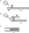
Models of replication control for γ ori. The primase-occluding model (A and B) and the handcuffing model (C) are shown. See text and reference for details.

Similar articles

Cited by

References

    1. Bouche J P, Rowen L, Kornberg A. The RNA primer synthesized by primase to initiate phage G4 DNA replication. J Biol Chem. 1978;253:765–769. - PubMed
    1. Bramhill D, Kornberg A. Duplex opening by DnaA protein at novel sequences in initiation of replication at the origin of the E. coli chromosome. Cell. 1988;52:743–755. - PubMed
    1. Chen D, Feng J, Kruger R, Urh M, Inman R B, Filutowicz M. Replication of R6K γ origin in vitro: discrete start sites for DNA synthesis dependent on π and its copy-up variants. J Mol Biol. 1998;282:775–787. - PubMed
    1. Dellis S, Feng J, Filutowicz M. Replication of plasmid R6K γ origin in vivo and in vitro: dependence on IHF binding to the ihf1 site. J Mol Biol. 1996;257:550–560. - PubMed
    1. Dellis S, Filutowicz M. Integration host factor of Escherichia coli reverses the inhibition of R6K plasmid replication by π initiator protein. J Bacteriol. 1991;173:1279–1286. - PMC - PubMed

Publication types