. 2000 Apr;81(2):117-43.
doi: 10.1046/j.1365-2613.2000.00146.x.

Epithelial stem cell repertoire in the gut: clues to the origin of cell lineages, proliferative units and cancer

Affiliations

Epithelial stem cell repertoire in the gut: clues to the origin of cell lineages, proliferative units and cancer

N A Wright. Int J Exp Pathol. 2000 Apr.

Abstract

Gastrointestinal stem cells are shown to be pluripotential and to give rise to all cell lineages in the epithelium. After damage, gut stem cells produce reparative cell lineages that produce a wide range of peptides with important actions on cell proliferation and migration, and promote regeneration and healing. Increase in stem cell number is considered to induce crypt fission, and lead to increases in the number of crypts, even in the adult; it is also the mode of spread of mutated clones in the colorectal mucosa. Stem cell repertoire is defined by both intrinsic programming of the stem cell itself, but signalling from the mesenchyme is also vitally important for defining both stem cell progeny and proliferation. Carcinogenesis in the colon occurs through sequential mutations, possibly occurring in a single cell. A case is made for this being the stem cell, but recent studies indicate that several stem cells may need to be so involved, since early lesions appear to be polyclonal in derivation.


Figures
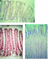
Figure 1
Figure 1
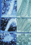
The histological appearances and organization of (a) the colon, showing the crypts containing numerous mucus-secreting goblet cells; (b) the small intestine, with the surface villi receiving cells migrating from the crypts and (c) the acid-secreting gastric mucosa. Here the surface and foveolar cells are mucus-secreting, while deeper in the glands the large parietal cells and the more darkly staining peptic or chief cells are prominent. The mucous neck cells are found in the neck region, where they appear as triangular, mucus-secreting cells sitting between the parietal cells.
Figure 2
Figure 2
A classification of cells in a renewal system. The stem cells form a self-renewing population, providing dividing transit cells, of limited proliferative potential, which, after a small number of divisions, decycle and begin the differentiation process proper.
Figure 3
Figure 3
A diagrammatic representation of the Unitarian Theory of Cytogenesis in (a) the intestine (courtesy of Professor C.P. Leblond); (b) a lineage diagram for the production of columnar cells (C) and mucous cells (M), generated by the stem cell (S) based on clonal analysis of Dlb-1 positive clones in SWR mice. Large arrows indicate dominant pathways. Mix is a short-lived progenitor capable of producing both C and M cell precursors (C1 and M1). C0 and M0 are long-lived precursors. C1 and M1 can be amplified by transit divisions (reproduced from Bjerknes & Cheng (1999), with permission); (c) the gastric fundic gland. The crypt-base columnar stem cells in the intestinal crypt give rise to the cell lineages shown while undifferentiated granule-free cells in the isthmus of the gastric gland are regarded as stem cells and give origin to the three committed precursors. In the gastric gland there is a bi-directional flux of cells, upwards to the surface (the presurface or prepit cells giving rise to the surface or pit cells), and downwards upwards the base of the gland (parietal, chief, ECL and other endocrine cells). The mucous neck cells remain in the neck (redrawn from Karam & Leblond 1993).
Figure 4
Figure 4
The Pearse hypothesis, in which neuroendocrine-programmed stem cells from the primitive epiblast colonize the gut and pancreatic primordia (courtesy of Professor A.G.E. Pearse and Churchill Livingstone).
Figure 5
Figure 5
An endocrine cell, immunostained with chromogranin A, growing in a culture of HRA-19 cells; note the typical bipolar morphology (courtesy of Dr Susan Kirkland).
Figure 6
Figure 6
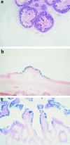
(a) section of a tumour grown from the single-cell cloned HRA-19a cell line in the flank of a nude mouse; (b) from a xenograft of the HRA-19a single cell cloned cell line growing in a nude mouse, demonstrating the presence of goblet cells stained with Alcian Blue (courtesy Dr Susan Kirkland).
Figure 7
Figure 7
Endocrine cells in the HRA-19a xenograft demonstrated by (a) the Grimelius technique, and (b) by immunostaining with chromogranin A (courtesy of Dr Susan Kirkland).
Figure 8
Figure 8
Section from the jejunal mucosa and colon of a tetraparental allophenic mouse stained with the lectin Dolichos biflorus agglutinin, which differentiates between the two types of cells present in the chimaera. Note in (a), and confirm in (b) (at high power), that crypts are either totally positive or totally negative, indicating that each crypt was originally the progeny of a single cell, or group of cells, of the same type, and supporting the concept of a clonal origin of crypts. Effectively similar results are seen in the colonic crypts (c). The polymorphism is also shown in the endothelial cells (photographed from a preparation made available through the courtesy of Professor B Ponder).
Figure 9
Figure 9
A section from the colon of a mouse heterozygous for a defective glucose-6-phosphate dehydrogenase (G6PD) gene, stained for G6PD activity. Most of the mucosa is positive, but single crypts are wholly negative (courtesy of Professor Geraint Williams).
Figure 10
Figure 10
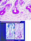
A low power view of a section of the stomach of an XX/XY chimaeric mouse, contemporaneously subjected to in situ hybridization to demonstrate the highly repetitive sequences in the mouse Y chromosome using digoxigenin: contiguous male and female areas are readily seen. (Courtesy of Dr Mary Thompson).
Figure 11
Figure 11
Sections from the colon of a mouse treated with a single injection of a mutagen (ethyl nitrosourea, ENU), and histochemically stained for glucose-6-phosphatase activity: (a) a partially negative crypt, where only a portion of the crypt is positive, and (b) a wholly negative crypt without any positive cells; (c) a patch of negative-staining cells (courtesy of Dr Hyun Sook Park).
Figure 12
Figure 12
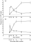
The rate of appearance in (a) small intestine and (b) colon of partially (▪,•) and wholly (□,○) negative crypts in mice treated with ethyl nitrosourea (□,▪) and control animals (○,•). Results are expressed as number per 104 crypts; animals were injected with ENU at six weeks of age (courtesy of Dr Hyun Sook Park).
Figure 13
Figure 13
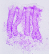
A section from the human colon stained with the mild periodic acid-Schiff method for nonacetylated sialomucin; note that the loss of acetylation is confined to single crypt — crypt restricted, and that all goblet cells in the crypt are negatively stained (courtesy of Dr Fiona Campbell).
Figure 14
Figure 14
The stomach of the XX/XY chimaeric mouse, contemporaneously subjected to in situ hybridization to demonstrate the highly repetitive sequences in the mouse Y chromosome using digoxigenin, and to show gastrin cells using an antigastrin antibody (a) a male area containing definitive male endocrine cells with blue dots; (b) a female area showing endocrine cells which do not bear a blue spot, indicating their female nature (courtesy of Dr Mary Thompson).
Figure 15
Figure 15
Sections from a patient with the XO/XY phenotype, with the XY cell demonstrated by in situ hybridization: (a) showing all cells positive or negative for the Y chromosome; (b) showing goblet cell clonality; (c) sections stained with chromogranin A for endocrine cells (red), showing clonal derivation; (d) showing a hemi-crypt which has lost nuclear staining for the Y chromosome (from Novelli et al. 1996, with permission).
Figure 16
Figure 16
(a) showing the early buds of the UACL growing out of parent crypts close to an ulcerated area of the mucosa in Crohn's disease. Note the abrupt origin of the UACL cells, from the stem cell zone, and the absence of mitotic activity in the UACL cells; (b) a mature UACL complex, showing the acinar area, the duct, and the UACL cells clothing the surface of a villus, displacing the indigenous cell lineages; note the single goblet cell in the duct area (c) a more developed acinar complex, growing as a newly formed gland in the lamina propria (Reproduced from Wright et al. 1990a, with permission).
Figure 17
Figure 17
A microdissected crypt, stained with Feulgen's reagent, showing the mode of crypt fission.
Figure 18
Figure 18
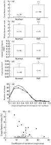
Comparisons of parameters in control mucosa and mucosa from patients with FAP. (a) the crypt fission index; (b) numbers of mitoses per crypt; (c) the position of the highest mitotic figure in the crypt; (d) the crypt area; (e) the distribution of mitoses in control (○, n = 40) and FAP mucosa (•, n = 40). (f) the relationship between the coefficient of variation of the crypt area (Cv) and the crypt fission index. Higher crypt fission indices were associated with larger Cvs.in normal mucosa (○, r = 0.44, n = 66, P = 0.002, and lower crypt fission indices with larger Cvs.in FAP mucosa (•, r = 0.68, n = 12, P = 0.005) (courtesy of Dr Hyun Sook Park).
Figure 19
Figure 19
A diagram showing the distribution of aneuploid stem lines in the colon of a patient with ulcerative colitis and dysplasia; note the large areas, particularly in the sigmoid colon and transverse colon, which have been colonized by the same stem cell line. From Levin et al. (1991), with permission.
Figure 20
Figure 20
(a) rat small intestine which has been transplanted subcutaneously into nude mice as a piece of developing gut endoderm embedded in collagen, and sectioned some 14 days later, stained with haematoxylin and eosin; (b) endoderm which has been transfected with the BAG retrovirus, showing expression of β-galactosidase; (c) at 6 weeks after transplantation. Note the presence of β-gal staining confined to one side of the villus, indicating that crypts stably transfected with the retrovirus are feeding only that side of the villus. This suggests that transfection is crypt-restricted, but that expression of β-gal is transcriptionally regulated at the level of the crypt-villus junction (courtesy of Dr R. Del Buono).
Figure 21
Figure 21
A colonic polyp from a Cdx 2+/− mouse showing colonic mucosa (C), small intestinal epithelium (SI), gastric antral (GA), and gastric corpus mucosa (GCo) with characteristic parietal cells (courtesy of Professor Felix Beck).
Figure 22
Figure 22
A microadenoma from the XO/XY patient, showing adenomatous crypts which are positive or negative for the Y chromosome in the same lesion, demonstrated in the lower part of the figure (from Novelli et al. 1996, with permission).

References

    1. Ahnen D, Gullick W, Wright NA. Expression of multiple growth factors by the ulcer-associated cell lineage. Gastroenterology. 1991;100:A512.
    1. Ahnen D. Abnormal DNA content as a biomarker of large bowel cancer risk and prognosis. J. Cell Biochem. 1992;16(Suppl.):143–150. - PubMed
    1. Ahnen D, Poulsom R, Stamp G, et al. The ulceration-associated cell lineage reiterates the differentiation programme of Brunners glands, but acquires the proliferative organisation of the gastric gland. J. Pathol. 1994;173:317–326. - PubMed
    1. Alexander P. Do cancers arise from a single transformed cell or is monoclonality of tumours a late event in carcinogenesis. Br. J. Cancer. 1985;51:453–457. - PMC - PubMed
    1. Andrew A. Further evidence that enterochromaffin cells are not derived from the neural crest. J. Embryol. Exp. Morph. 1973;31:581–598. - PubMed