Review
. 2000 Apr 28;298(2):167-85.
doi: 10.1006/jmbi.2000.3668.

Structure, stability and function of RNA pseudoknots involved in stimulating ribosomal frameshifting

Affiliations
Review

Structure, stability and function of RNA pseudoknots involved in stimulating ribosomal frameshifting

D P Giedroc et al. J Mol Biol. .

Abstract

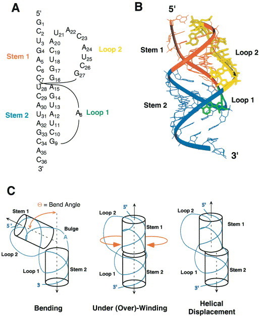
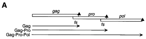
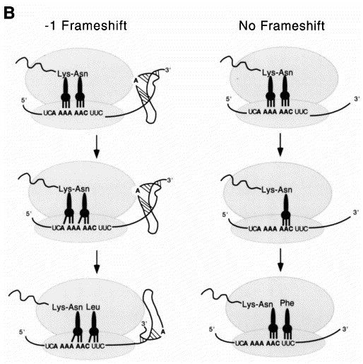
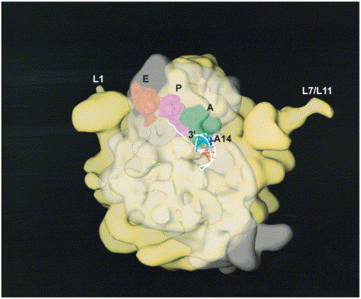
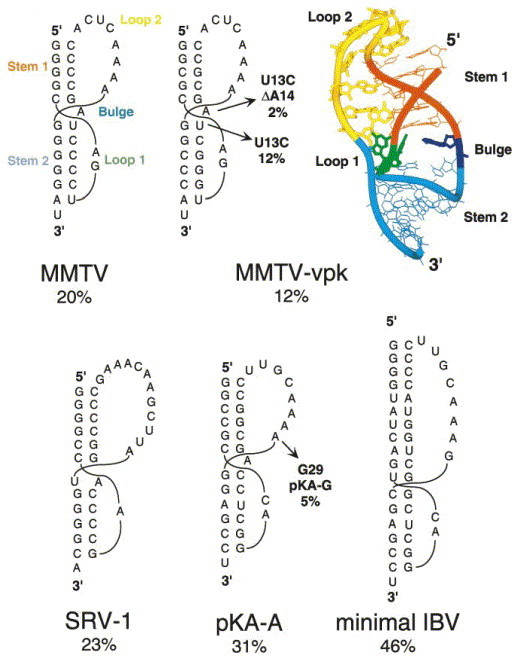
Programmed -1 ribosomal frameshifting has become the subject of increasing interest over the last several years, due in part to the ubiquitous nature of this translational recoding mechanism in pathogenic animal and plant viruses. All cis-acting frameshift signals encoded in mRNAs are minimally composed of two functional elements: a heptanucleotide "slippery sequence" conforming to the general form X XXY YYZ, followed by an RNA structural element, usually an H-type RNA pseudoknot, positioned an optimal number of nucleotides (5 to 9) downstream. The slippery sequence itself promotes a low level ( approximately 1 %) of frameshifting; however, downstream pseudoknots stimulate this process significantly, in some cases up to 30 to 50 %. Although the precise molecular mechanism of stimulation of frameshifting remains poorly understood, significant advances have been made in our knowledge of the three-dimensional structures, thermodynamics of folding, and functional determinants of stimulatory RNA pseudoknots derived from the study of several well-characterized frameshift signals. These studies are summarized here and provide new insights into the structural requirements and mechanism of programmed -1 ribosomal frameshifting.


Figures

Figure 1
Figure 1
(a) Secondary structural representation of an H-type pseudoknot. The sequence shown corresponds to the autoregulatory gene 32 mRNA pseudoknot from bacteriophage T2 (Du et al., 1996). (b) Tertiary structural representation of the simple pseudoknotted RNA from the T2 gene 32 mRNA showing that the single-stranded loops L1 (green) and L2 (yellow) will cross the major and minor grooves of stems S2 (blue) and S1 (red), respectively, on the same face of the molecule provided the junction region conforms to near-normal A-form geometry (PDB code 2TPK). (c) Schematic representation of the structural diversity of the helical junction of simple H-type RNA pseudoknots. Note that various circular permutations of this motif can theoretically be formed, e.g. by breaking the chain in loop L1 or L2 and connecting the 5′ and 3′ ends shown with an extended loop. The standard H-type folding topology shown is nearly exclusively observed in natural RNA sequence contexts.
Figure 2
Figure 2
(a) Production of Gag, Gag-Pro, and Gag-Pro-Pol fusion proteins from a single mRNA by way of two isolated −1 ribosomal frameshifting events. (b) Simultaneous slippage model (Jacks et al., 1988a) for translational regulation of ribosomal frameshifting. Adapted from Gesteland & Atkins (1996).
Figure 2
Figure 2
(a) Production of Gag, Gag-Pro, and Gag-Pro-Pol fusion proteins from a single mRNA by way of two isolated −1 ribosomal frameshifting events. (b) Simultaneous slippage model (Jacks et al., 1988a) for translational regulation of ribosomal frameshifting. Adapted from Gesteland & Atkins (1996).
Figure 3
Figure 3
Model of the MMTV gag-pro mRNA frameshifting signal docked into the low resolution structure of the E. coli ribosome derived from the 25 Å cryo-electron microscopy electron density map (Frank et al., 1995a). The large 50 S ribosomal subunit (yellow) is shown to the rear of this view with the L1 stalk and the L7/L12 regions labeled. The small 30 S subunit is shown to the front, but made transparent so as not to obscure the aminoacyl (A), peptidyl (P) and exit (E) tRNAs, as well as the proposed path of the mRNA (Frank et al., 1995a). The A, P, and E-site tRNAs were positioned as described in the 7.8 Å resolution crystallographic structure of the T. thermophilus ribosome (Cate et al., 1999) (PDB code 486D) by manually docking them into the structure of the E. coli ribosome. The nucleotides of the slippery sequence are positioned opposite the A- and P-site tRNA anticodon triplets in the zero reading frame as described in the Cate et al. (1999) structure. The seven nucleotides between the 3′ base of the slippery sequence and the 5′ nucleotide of stem S1 are drawn in an extended conformation. The structure suggests that the pseudoknot need not be drawn into the ribosome and/or become largely unfolded when the slippery sequence is positioned in the active site of the ribosome. The structure further reveals how the 5′-side of loop L2 (as well as the base of stem S1) may well be first to physically interact with the translating ribosome. Adapted from Frank et al. (1995b).
Figure 4
Figure 4
Secondary structural representations and reported in vitro frameshifting efficiencies of the MMTV and MMTV-vpk gag-pro pseudoknots Chamorro et al 1992, Chen et al 1995, the SRV-1 gag-pro pseudoknot ten Dam et al 1994, Du et al 1997, and the minimal wild-type IBV (Liphardt et al., 1999) and chimeric MMTV-IBV (pKA-A) (Napthine et al., 1999) pseudoknots. The solution structure of the MMTV-vpk pseudoknot is also shown (PDB code 1RNK) (Shen & Tinoco, 1995).
Figure 5
Figure 5
(a) The structure of the 28-nucleotide BWYV pseudoknot as determined by X-ray crystallography (Su et al., 1999) (PDB code 437D). The C8·G12-C26 base-triple interaction is shown in red and the loop 2 nucleotides that make crystal contacts with stem 1 are in blue. Also shown are U13 and A25 which would be predicted to form a base-pair in stem S2 (green) and a Na+ ion bound between loop L2 and stem S1 nucleotides (yellow). (b) Secondary structural rendering of the BWYV pseudoknot with nucleotides colored as in (a).
Figure 6
Figure 6
(a) The equilibrium unfolding pathway of the bacteriophage T4 gene 32 autoregulatory mRNA pseudoknot. q is the partition function for this coupled equilibrium with the folded pseudoknot as the reference state. Derived from Theimer et al. (1998). (b) Cartoon representations of two possible unfolding pathways which might occur at the ribosome.
Figure 6
Figure 6
(a) The equilibrium unfolding pathway of the bacteriophage T4 gene 32 autoregulatory mRNA pseudoknot. q is the partition function for this coupled equilibrium with the folded pseudoknot as the reference state. Derived from Theimer et al. (1998). (b) Cartoon representations of two possible unfolding pathways which might occur at the ribosome.
Figure 7
Figure 7
Hypothetical reaction coordinate diagrams which illustrate thermodynamic (a) versus primarily kinetic (b) control of partial unfolding of the pseudoknot. F, folded; hp, hairpin intermediate; U, unfolded RNAs schematized for a wild-type, functional pseudoknot (—) and a weakly destabilized mutant pseudoknot (– – –). In (a), the transition state free energies are identical; thus ΔΔGF↔hp is totally determined by ΔΔGF↔hp or the ground-state structure. In (b), a small ΔΔGF↔hp is shown accompanied by a significant difference in GF↔hp for each of two RNAs such that ΔΔGF↔hp are different. In both cases, a more positive ΔGF↔hp for the functional pseudoknot would slow the rate of interconversion between the F and partially unfolded forms, potentially increasing the efficiency of frameshifting (see the text for details).

References

    1. Akke M, Fiala R, Jiang F, Patel D, Palmer A.G.I. Base dynamics in a UUCG tetraloop RNA hairpin characterized by 15N spin relaxation: Correlations with structure and stability. RNA. 1997;3:702–709. - PMC - PubMed
    1. Alam S.L, Atkins J.F, Gesteland R.F. Programmed ribosomal frameshifting: much ado about knotting! Proc. Natl Acad. Sci. USA. 1999;96:14177–14179. - PMC - PubMed
    1. Alam S.L, Wills N.M, Ingram J.A, Atkins J.F, Gesteland R.F. Structural studies of the RNA pseudoknot required for readthrough of the gag-termination codon of murine leukemia virus. J. Mol. Biol. 1999;288:837–852. - PubMed
    1. Atkins J.F, Weiss R.B, Gesteland R.F. Ribosome gymnastics: degree of difficulty 9.5, style 10.0. Cell. 1990;62:413–423. - PMC - PubMed
    1. Ban N, Nissen P, Hansen J, Capel M, Moore P.B, Steitz T.A. Placement of protein and RNA structures into a 5 Å-resolution map of the 50 S ribosomal subunit. Nature. 1999;400:841–847. - PubMed

Publication types

MeSH terms

LinkOut - more resources