. 2000 Apr 1;14(7):854-62.

Severe liver degeneration and lack of NF-kappaB activation in NEMO/IKKgamma-deficient mice

Affiliations

Severe liver degeneration and lack of NF-kappaB activation in NEMO/IKKgamma-deficient mice

D Rudolph et al. Genes Dev. .

Abstract

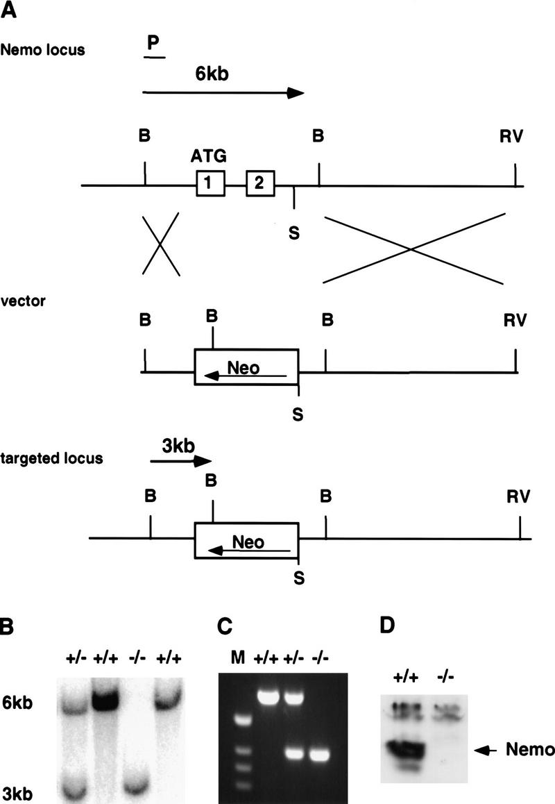
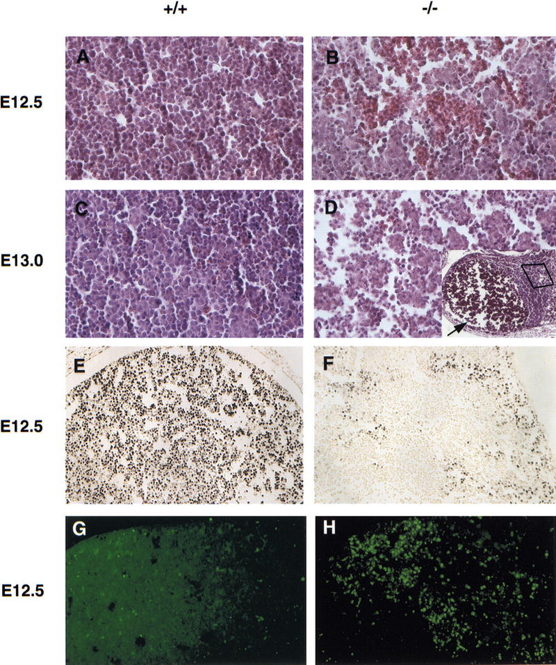
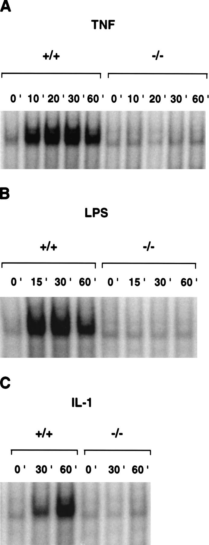
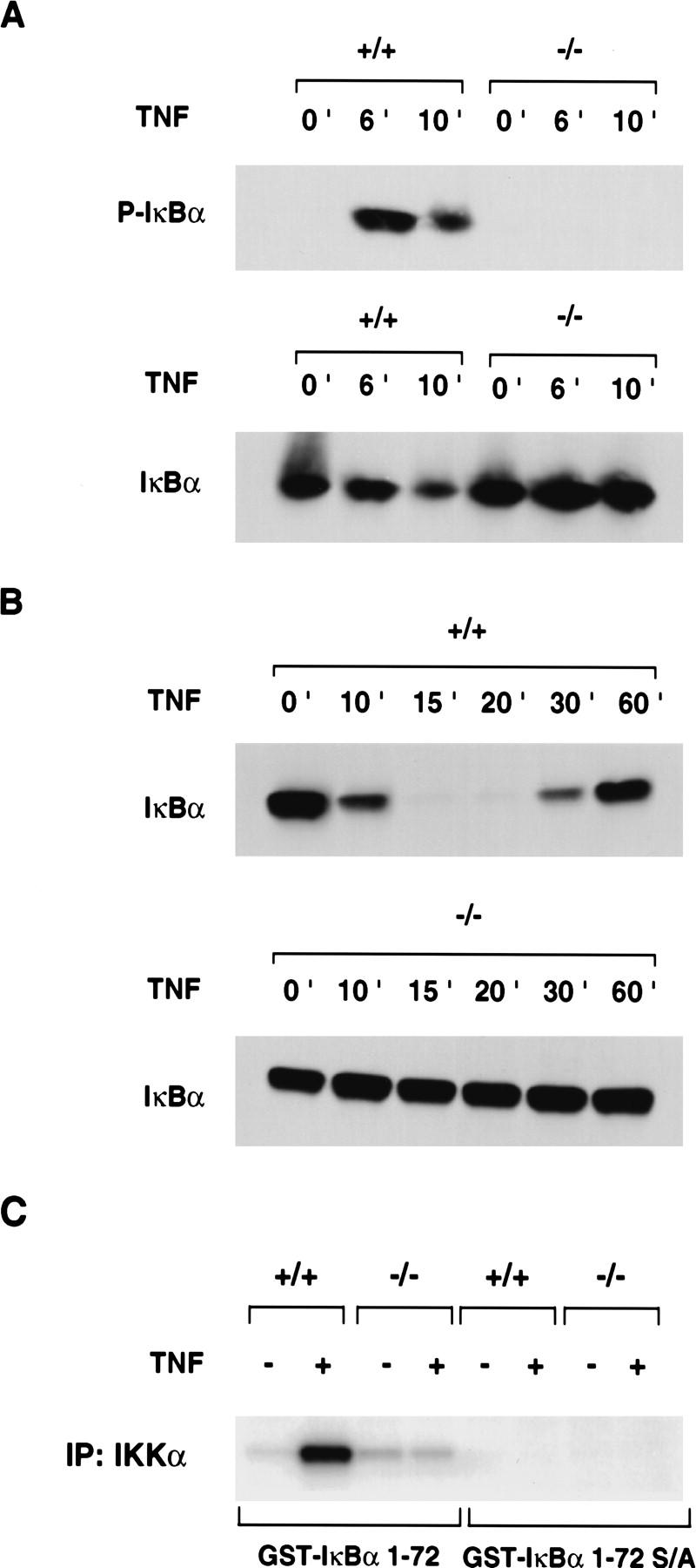
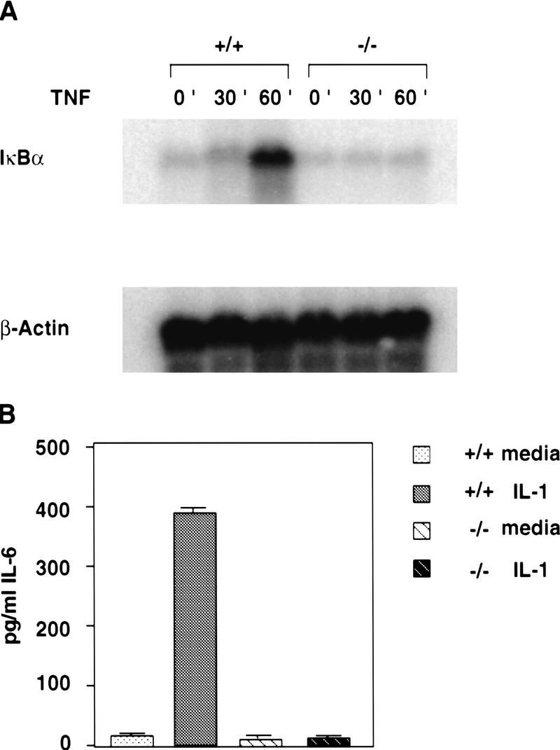
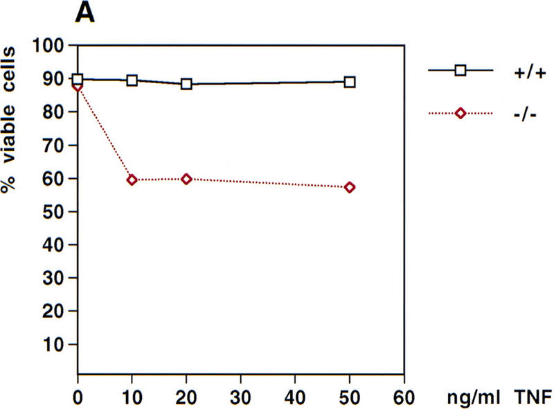
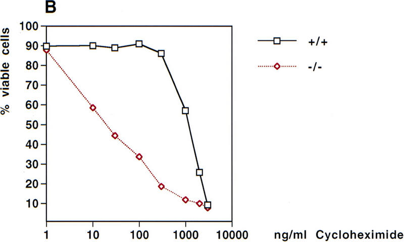
Phosphorylation of IkappaB, an inhibitor of NF-kappaB, is an important step in the activation of the transcription factor NF-kappaB. Phosphorylation is mediated by the IkappaB kinase (IKK) complex, known to contain two catalytic subunits: IKKalpha and IKKbeta. A novel, noncatalytic component of this kinase complex called NEMO (NF-kappaB essential modulator)/IKKgamma was identified recently. We have generated NEMO/IKKgamma-deficient mice by gene targeting. Mutant embryos die at E12.5-E13.0 from severe liver damage due to apoptosis. NEMO/IKKgamma-deficient primary murine embryonic fibroblasts (MEFs) lack detectable NF-kappaB DNA-binding activity in response to TNFalpha, IL-1, LPS, and Poly(IC) and do not show stimulus-dependent IkappaB kinase activity, which correlates with a lack of phosphorylation and degradation of IkappaBalpha. Consistent with these data, mutant MEFs show increased sensitivity to TNFalpha-induced apoptosis. Our data provide in vivo evidence that NEMO/IKKgamma is the first essential, noncatalytic component of the IKK complex.


Figures

Figure 1
Figure 1
Generation of NEMO/IKKγ−/− mice. (A) Wild-type NEMO/IKKγ locus (top). Exon 1 and 2 are shown as open boxes and the ATG translation start site is indicated. (P) External flanking probe, (B) BamHI, (RV) EcoRV, (S) SpeI. The targeting vector (middle) was designed to replace exon 1 and exon 2 of the wild-type locus with a neomycin cassette in antisense orientation. Flanking probe P detects a 6-kb BamHI fragment corresponding to the wild-type allele and a 3-kb BamHI fragment for the targeted allele (bottom). (B) Genomic Southern blot of BamHI-digested DNA from E12.5 embryos of the indicated genotypes, showing the 6-kb wild-type band and the 3-kb mutant band detected by probe P. (C) Genotyping by PCR of DNA from E12.5 embryos using the primers described in Material and Methods. (D) Western blot analysis of NEMO/IKKγ protein expression in cytoplasmic extracts of MEFs of the indicated genotypes.
Figure 2
Figure 2
Massive liver apoptosis in the absence of NEMO/IKKγ. (A–D) Histological analysis of liver sections from E12.5 (A,B) and E13.0 (C,D) wild-type (A,C) and NEMO/IKKγ-deficient (B,D) embryos. Hematoxylin and eosin staining; high-power view. (D) The inset shows a low power view of the same region shown in D. The arrow points to a large area of tissue loss in which erythrocytes have accumulated. (E,F) Detection of cell proliferation as determined by BrdU incorporation. Pregnant females were injected IP with BrdU 60-min prior to sacrifice. Sections of E12.5 wild-type and mutant embryos were prepared and immunostained with anti-BrdU antibody. A high power view of the liver is shown. (G,H) Enhanced apoptosis in livers of E12.5 NEMO/IKKγ-deficient embryos. TUNEL assays were performed on sections of wild-type and mutant E12.5 embryos. A high power view of the liver is shown in both cases.
Figure 3
Figure 3
Impaired NF-κB activation in NEMO/IKKγ-deficient cells. Wild-type or NEMO/IKKγ−/− MEFs were incubated with mouse recombinant TNFα (10 ng/ml) (A), LPS (15 μg/ml) (B), or IL-1 (10 ng/ml) (C) for the indicated times. NF-κB activation in 10 μg of nuclear extract was determined by gel shift assay as described in Materials and Methods.
Figure 4
Figure 4
Defective TNFα-induced IκBα phosphorylation and degradation and IκB kinase activity in the absence of NEMO/IKKγ. (A) IκBα phosphorylation. (Top) Wild-type and NEMO/IKKγ-deficient MEFs were treated with TNFα (10 ng/ml) for the indicated times followed by Western blot analysis of cytoplasmic extracts with anti-phospho-IκBα-specific antibody. (Bottom) Western blot of the same extracts with anti-IκBα antibody as a control to show IκBα protein expression in mutant cells. (B) IκBα degradation. Wild-type (top) and NEMO/IKKγ-deficient (bottom) MEFs were treated with TNFα (10 ng/ml) for the indicated times followed by Western blot analysis with anti-IκBα specific antibody. (C) IκB kinase activity. Wild-type and NEMO/IKKγ-deficient MEFs were either left in medium alone or treated with TNFα (10 ng/ml) for 7 min. Whole cell extracts were immunoprecipitated with anti-IKKα antibody and in vitro kinase activity was determined as described in Materials and Methods using GST–IκBα (1–72) (left) or GST–IκBα (1–72) S/A (right) as a substrate. One representative experiment of four is shown.
Figure 5
Figure 5
Impaired expression of NF-κB target genes in NEMO/IKKγ-deficient MEFs. (A) TNFα-induced IκBα mRNA synthesis. Wild-type and NEMO/IKKγ-deficient MEFs were stimulated with TNFα (10 ng/ml) for the indicated times and 20 μg of RNA was analyzed by Northern blot analysis using a 32P-labeled random priming probe corresponding to the full-length IκBα cDNA. β-actin, loading control. (B) IL-1 induced IL-6 production. Wild-type and NEMO/IKKγ-deficient MEFs (3 × 104) were left untreated or stimulated with IL-1 (10 ng/ml) for 16 hr. Levels of IL-6 (pg/ml) in culture supernatants were determined by ELISA. Values shown are the mean and standard deviation of triplicate samples.
Figure 6
Figure 6
TNFα-induced cytotoxicity. (A) TNFα-induced cytotoxicity. Wild-type and NEMO/IKKγ-deficient (2 × 105) MEFs were left untreated or stimulated with the indicated concentrations of TNFα for 18 hr. Cells were then harvested and stained with PI/AnnexinV-FITC. Values shown are the percentage of viable cells after treatment relative to untreated controls. (B) Cytotoxicity induced by TNFα plus cycloheximide. Wild-type and NEMO/IKKγ-deficient MEFs (2 × 105) were left untreated or stimulated with TNFα (10 ng/ml) in combination with the indicated concentrations of cycloheximide for 18 hr. Cells were harvested and stained as described for A, and values shown were determined as for A.
Figure 6
Figure 6
TNFα-induced cytotoxicity. (A) TNFα-induced cytotoxicity. Wild-type and NEMO/IKKγ-deficient (2 × 105) MEFs were left untreated or stimulated with the indicated concentrations of TNFα for 18 hr. Cells were then harvested and stained with PI/AnnexinV-FITC. Values shown are the percentage of viable cells after treatment relative to untreated controls. (B) Cytotoxicity induced by TNFα plus cycloheximide. Wild-type and NEMO/IKKγ-deficient MEFs (2 × 105) were left untreated or stimulated with TNFα (10 ng/ml) in combination with the indicated concentrations of cycloheximide for 18 hr. Cells were harvested and stained as described for A, and values shown were determined as for A.

References

    1. Akira S, Kishimoto T. IL-6 and NF-IL6 in acute-phase response and viral infection. Immunol Rev. 1992;127:25–50. - PubMed
    1. Baeuerle PA, Henkel T. Function and activation of NF-κB in the immune system. Annu Rev Immunol. 1994;12:141–179. - PubMed
    1. Beg AA, Baltimore D. An essential role for NF-κB in preventing TNF-α-induced cell death. Science. 1996;274:782–784. - PubMed
    1. Beg AA, Sha WC, Bronson RT, Ghosh S, Baltimore D. Embryonic lethality and liver degeneration in mice lacking the RelA component of NF-κB. Nature. 1995;376:167–170. - PubMed
    1. Cohen L, Henzel WJ, Baeuerle PA. IKAP is a scaffold protein of the IκB kinase complex. Nature. 1998;395:292–296. - PubMed

Publication types

LinkOut - more resources