

Heat production in human skeletal muscle at the onset of intense dynamic exercise
- PMID: 10766936
- PMCID: PMC2269891
- DOI: 10.1111/j.1469-7793.2000.00603.x
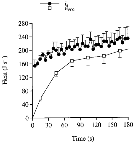
Heat production in human skeletal muscle at the onset of intense dynamic exercise
Abstract
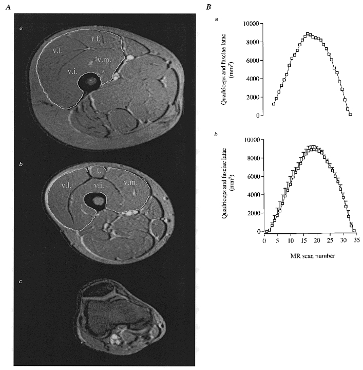
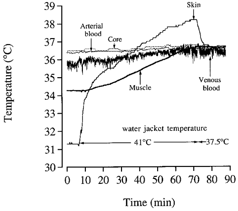
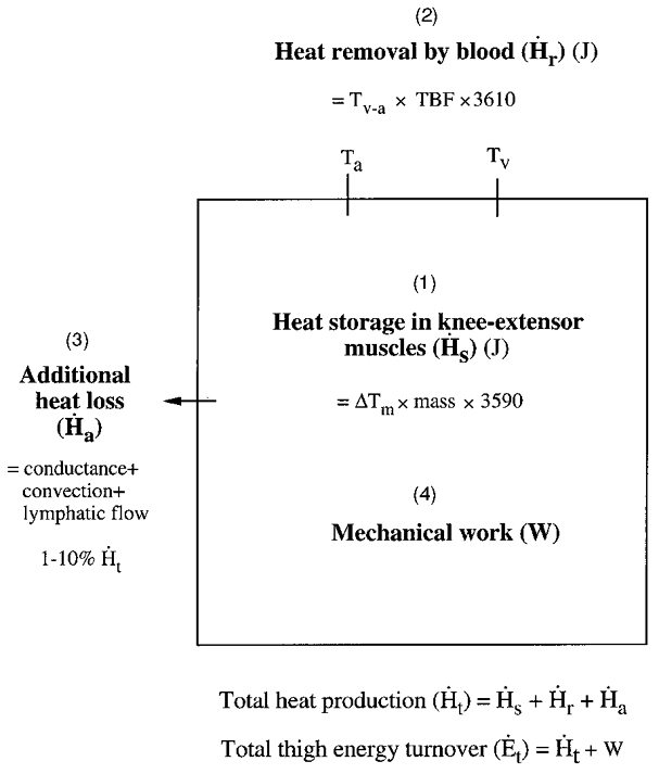
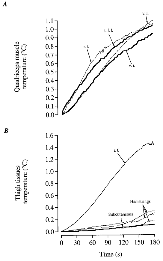
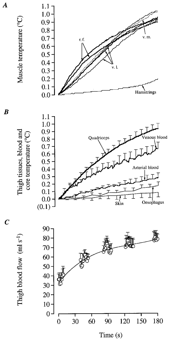
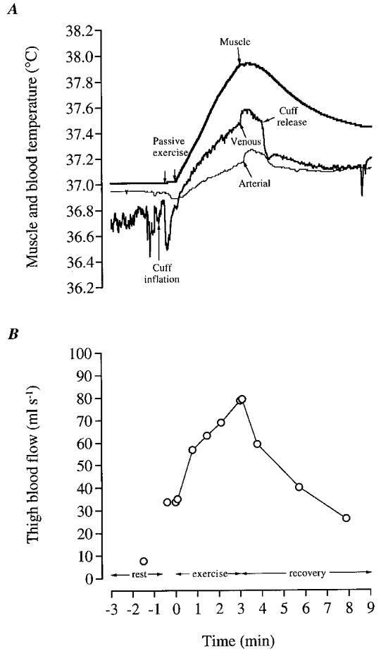
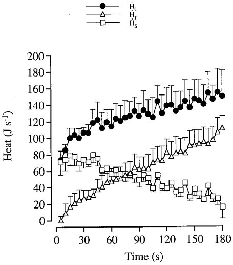
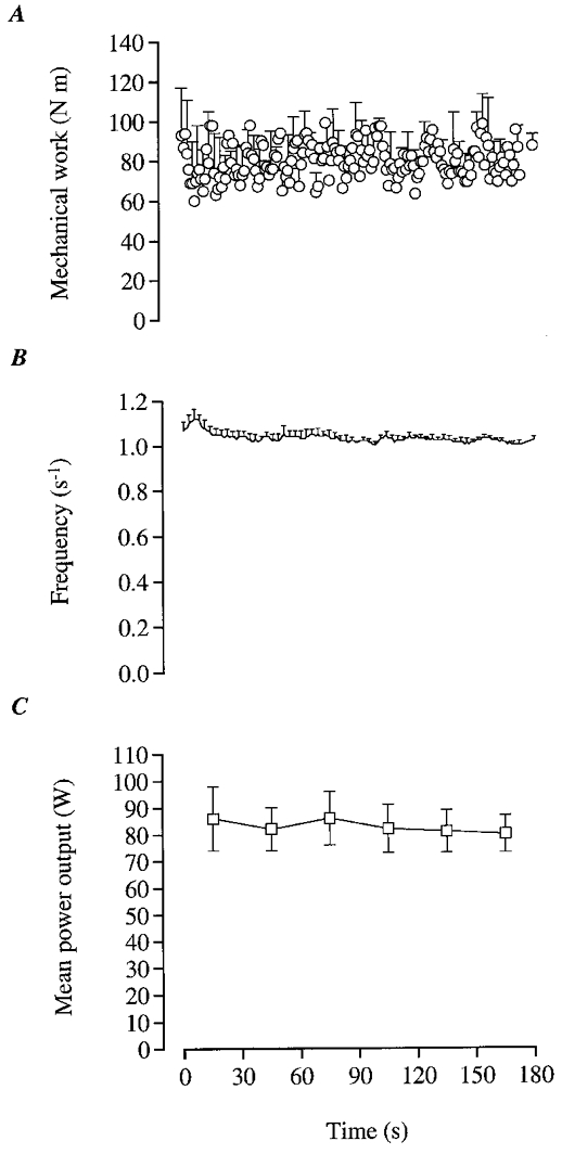
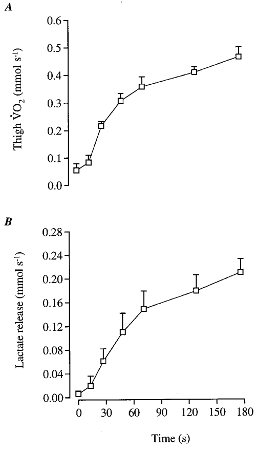
1. We hypothesised that heat production of human skeletal muscle at a given high power output would gradually increase as heat liberation per mole of ATP produced rises when energy is derived from oxidation compared to phosphocreatine (PCr) breakdown and glycogenolysis. 2. Five young volunteers performed 180 s of intense dynamic knee-extensor exercise ( approximately 80 W) while estimates of muscle heat production, power output, oxygen uptake, lactate release, lactate accumulation and ATP and PCr hydrolysis were made. Heat production was determined continuously by (i) measuring heat storage in the contracting muscles, (ii) measuring heat removal to the body core by the circulation, and (iii) estimating heat transfer to the skin by convection and conductance as well as to the body core by lymph drainage. 3. The rate of heat storage in knee-extensor muscles was highest during the first 45 s of exercise (70-80 J s-1) and declined gradually to 14 +/- 10 J s-1 at 180 s. 4. The rate of heat removal by blood was negligible during the first 10 s of exercise, rising gradually to 112 +/- 14 J s-1 at 180 s. The estimated rate of heat release to skin and heat removal via lymph flow was < 2 J s-1 during the first 5 s and increased progressively to 24 +/- 1 J s-1 at 180 s. The rate of heat production increased significantly throughout exercise, being 107 % higher at 180 s compared to the initial 5 s, with half of the increase occurring during the first 38 s, while power output remained essentially constant. 5. The contribution of muscle oxygen uptake and net lactate release to total energy turnover increased curvilinearly from 32 % and 2 %, respectively, during the first 30 s to 86 % and 8 %, respectively, during the last 30 s of exercise. The combined energy contribution from net ATP hydrolysis, net PCr hydrolysis and muscle lactate accumulation is estimated to decline from 37 % to 3 % comparing the same time intervals. 6. The magnitude and rate of elevation in heat production by human skeletal muscle during exercise in vivo could be the result of the enhanced heat liberation during ATP production when aerobic metabolism gradually becomes dominant after PCr and glycogenolysis have initially provided most of the energy.
Figures
Similar articles
-
Muscle heat production and anaerobic energy turnover during repeated intense dynamic exercise in humans.J Physiol. 2001 Nov 1;536(Pt 3):947-56. doi: 10.1111/j.1469-7793.2001.00947.x. J Physiol. 2001. PMID: 11691886 Free PMC article. Clinical Trial.
-
Muscle oxygen uptake and energy turnover during dynamic exercise at different contraction frequencies in humans.J Physiol. 2001 Oct 1;536(Pt 1):261-71. doi: 10.1111/j.1469-7793.2001.00261.x. J Physiol. 2001. PMID: 11579174 Free PMC article.
-
Effect of temperature on skeletal muscle energy turnover during dynamic knee-extensor exercise in humans.J Appl Physiol (1985). 2006 Jul;101(1):47-52. doi: 10.1152/japplphysiol.01490.2005. Epub 2006 Mar 2. J Appl Physiol (1985). 2006. PMID: 16514001
-
Energy system interaction and relative contribution during maximal exercise.Sports Med. 2001;31(10):725-41. doi: 10.2165/00007256-200131100-00003. Sports Med. 2001. PMID: 11547894 Review.
-
Oxygen uptake kinetics: old and recent lessons from experiments on isolated muscle in situ.Eur J Appl Physiol. 2003 Oct;90(3-4):242-9. doi: 10.1007/s00421-003-0994-0. Epub 2003 Oct 11. Eur J Appl Physiol. 2003. PMID: 14556076 Review.
Cited by
-
Effect of Handgrip Training in Extreme Heat on the Development of Handgrip Maximal Isometric Strength among Young Males.Int J Environ Res Public Health. 2021 May 14;18(10):5240. doi: 10.3390/ijerph18105240. Int J Environ Res Public Health. 2021. PMID: 34069110 Free PMC article.
-
Recruitment of fibre types and quadriceps muscle portions during repeated, intense knee-extensor exercise in humans.Pflugers Arch. 2004 Oct;449(1):56-65. doi: 10.1007/s00424-004-1304-3. Pflugers Arch. 2004. PMID: 15290298
-
Role of {alpha}1-adrenergic vasoconstriction in the regulation of skeletal muscle blood flow with advancing age.Am J Physiol Heart Circ Physiol. 2009 Feb;296(2):H497-504. doi: 10.1152/ajpheart.01016.2008. Epub 2008 Dec 5. Am J Physiol Heart Circ Physiol. 2009. PMID: 19060122 Free PMC article.
-
Sports and environmental temperature: From warming-up to heating-up.Temperature (Austin). 2017 Aug 4;4(3):227-257. doi: 10.1080/23328940.2017.1356427. eCollection 2017. Temperature (Austin). 2017. PMID: 28944269 Free PMC article. Review.
-
Methods for improving thermal tolerance in military personnel prior to deployment.Mil Med Res. 2020 Nov 29;7(1):58. doi: 10.1186/s40779-020-00287-z. Mil Med Res. 2020. PMID: 33248459 Free PMC article. Review.
References
-
- Aagaard P, Simonsen EB, Trolle M, Bangsbo J, Klausen K. Moment and power generation during maximal knee extensions performed at low and high speeds. European Journal of Applied Physiology. 1994;69:376–381. - PubMed
-
- Andersen P, Adams RP, Sjøgaard G, Thorboe A, Saltin B. Dynamic knee extension as model for study of isolated exercising muscle in humans. Journal of Applied Physiology. 1985;59:1647–1653. - PubMed
-
- Ardevol A, Adan C, Remesar X, Fernández-López JA, Alemany M. Hind leg heat balance in obese Zucker rats during exercise. Pflügers Archiv. 1998;435:454–464. - PubMed
-
- Åstrand I. Aerobic work capacity in men and women with special reference to age. Acta Physiologica Scandinavica. 1960;49(suppl. 169):67–158. - PubMed
Publication types
MeSH terms
Substances
LinkOut - more resources
Full Text Sources
Other Literature Sources
Medical