

Human papilloma virus in melanoma biopsy specimens and its relation to melanoma progression
- PMID: 10767787
- PMCID: PMC1421053
- DOI: 10.1097/00000658-200005000-00006
Human papilloma virus in melanoma biopsy specimens and its relation to melanoma progression
Abstract
Objectives: To evaluate melanoma biopsy specimens for human papilloma virus (HPV) and determine the relation between the presence of HPV, in vitro growth, and clinical progression of melanoma in the patients from whom the biopsy specimens were derived.
Summary background data: Ultraviolet radiation from sun exposure appears to be the primary causal agent in the development of cutaneous melanoma. However, other agents, including HPV, as observed in different epithelial carcinomas, may also play a role in melanoma development and progression.
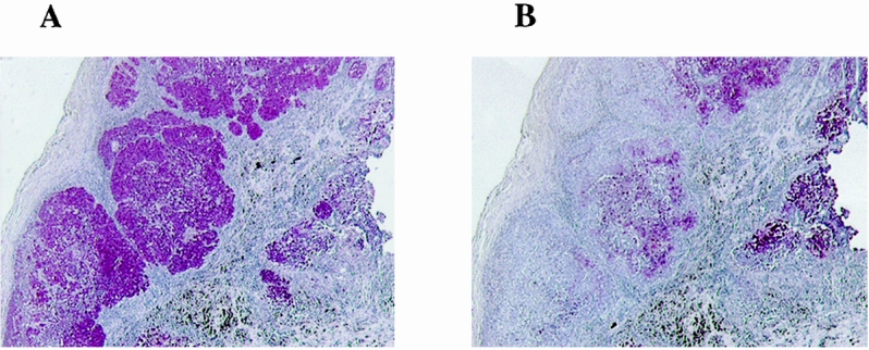
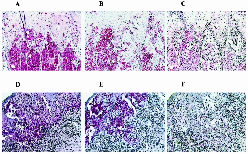
Methods: Twelve melanoma biopsy specimens obtained from 12 patients with AJCC stage III and IV melanoma were stained with antibodies against gp-100 (HMB-45) and S-100 protein to confirm melanoma diagnosis and with a polyclonal HPV antibody. After mechanical dissociation, the melanoma specimen cells' ability to grow in vitro was assessed. Patients were evaluated for melanoma progression with physical examination, complete blood count, and liver function tests every 3 months and a chest radiograph every 6 months.
Results: All biopsy specimens were positive for S-100, and nine (75%) were positive for gp-100. Seven of 12 (58%) were positive for HPV by immunohistochemistry. In vitro, none of the HPV-negative tumor cells grew from the tumor biopsies, whereas five of seven (71%) of the HPV-positive melanoma tumor cells grew very well. All patients with HPV-positive tumor cells had recurrences and died of melanoma progression, whereas four of five (80%) patients with HPV-negative tumor cells remained alive and without melanoma recurrence.
Conclusions: The presence of HPV was found in 58% of the biopsy specimens obtained from patients with stage III and IV melanoma and correlated with rapid melanoma progression. HPV may serve as a cofactor in the development of melanoma and may modulate a more aggressive phenotype in HPV-containing melanoma cells.
Figures
References
-
- Elder DE. Human melanocytic neoplasms and their etiologic relationship with sunlight. J Invest Dermatol 1989; 92:297S–303S. - PubMed
-
- Robinson JK, Rigel DS, Amonette RA. Trends in sun exposure knowledge, attitudes, and behaviors: 1986 to 1996. J Am Acad Dermatol 1997; 37:179–186. - PubMed
-
- van Elsas A, Scheibenbogen C, van der Minne C, Zerp SF, Keilholz U, Schrier PI. UV-induced N-ras mutations are T-cell targets in human melanoma. Melanoma Res 1997; 7:S107–S113. - PubMed
-
- Saenz-Santamaria MC, McNutt NS, Bogdany JK, Shea CR. p53 expression is rare in cutaneous melanomas. Am J Dermatopathol 1995; 17:344–349. - PubMed
-
- Swan DC, Vernon SD, Icenogle JP. Cellular proteins involved in papillomavirus-induced transformation. Arch Virol 1994; 138:105–115. - PubMed
Publication types
MeSH terms
LinkOut - more resources
Full Text Sources
Other Literature Sources
Medical
