

The importance of skip lesions in temporal arteritis
- PMID: 10767830
- PMCID: PMC1763281
- DOI: 10.1136/jcp.53.2.137
The importance of skip lesions in temporal arteritis
Abstract
Aim: To determine the frequency of skip lesions in an unselected series of temporal artery biopsies and compare the results with other series.
Methods: The study was a retrospective review of 102 consecutive temporal artery biopsies taken in a five year period (1992-1997) in one large hospital.
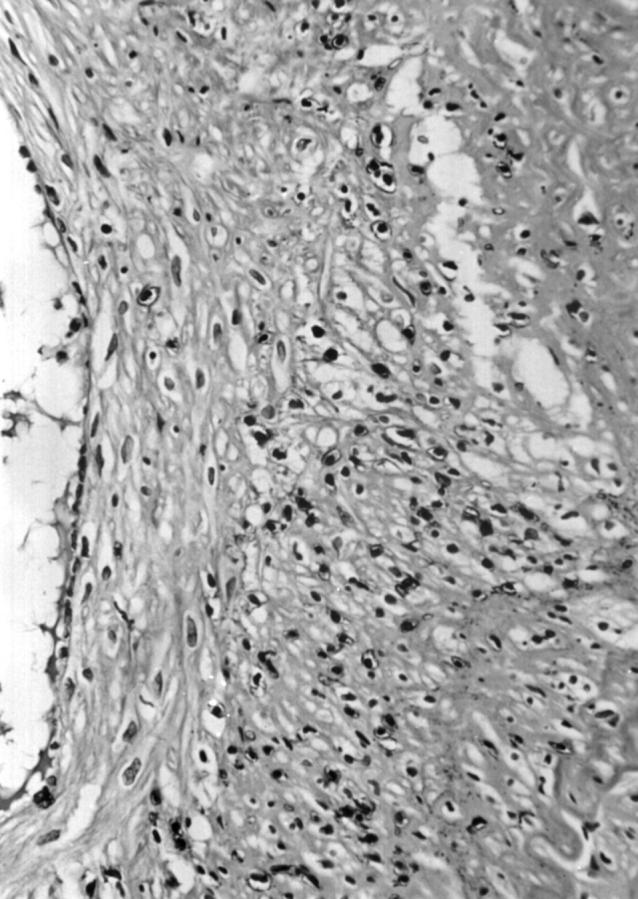
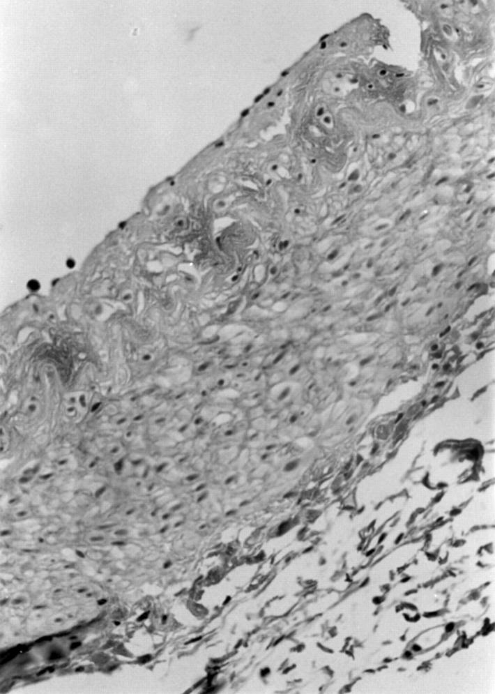
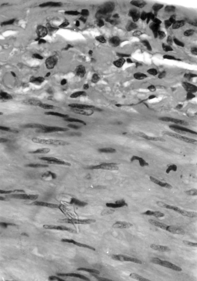
Results: 35 cases (34.3%) showed evidence of active cranial vasculitis with pathological evidence of inflammation of the intima or media, with or without giant cells. Three of these cases (8.5%) showed apparent skip lesions: normal intima, media, and adventitia in one segment while in other segments there was clear evidence of active vasculitis. Immunocytochemical stains for leucocyte common antigen (LCA) and CD15 were helpful in identifying the absence of intimal or medial inflammatory cell infiltrates within skip lesions. Skip lesions have been described in up to 28.3% of cases in some series, while others have not found evidence of skip lesions or have identified them in a much smaller percentage of cases.
Conclusions: In this series skip lesions were relatively rare, accounting for 8.5% of cases of active vasculitis. The degree of inflammation in temporal arteritis is discontinuous. Immunostaining for inflammatory cells, for example LCA and CD15, may be helpful in identifying the presence of an inflammatory cell infiltrate in skip lesion segments of the temporal artery.
Figures
References
MeSH terms
Substances
LinkOut - more resources
Full Text Sources
Medical
Research Materials