

Left atrial volume assessed by transthoracic three dimensional echocardiography and magnetic resonance imaging: dynamic changes during the heart cycle in children
- PMID: 10768903
- PMCID: PMC1760805
- DOI: 10.1136/heart.83.5.537
Left atrial volume assessed by transthoracic three dimensional echocardiography and magnetic resonance imaging: dynamic changes during the heart cycle in children
Abstract
Objective: To assess the dynamic changes in left atrial volume by transthoracic three dimensional echocardiography and compare the results with those obtained by magnetic resonance imaging (MRI).
Design and patients: 30 healthy children (15 boys and 15 girls, aged 8 to 13 years) underwent examination by three dimensional echocardiography and MRI.
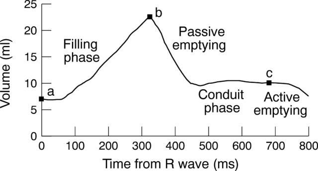
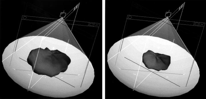
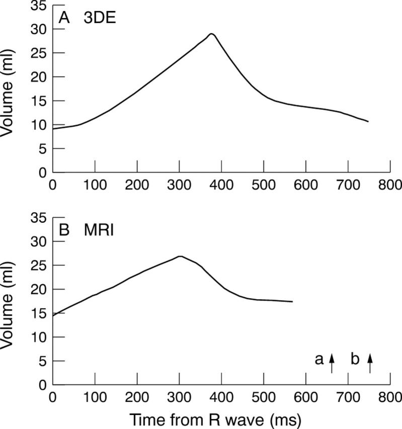
Methods: Three dimensional echocardiography of the left atrium was performed using rotational acquisition of planes at 18 degrees intervals from the parasternal window with ECG gating and without respiratory gating. Volume estimation by MRI was performed with a slice thickness of 4-8 mm and ECG triggering during breath holding in deep inspiration. A left atrial time-volume curve was reconstructed in each child.
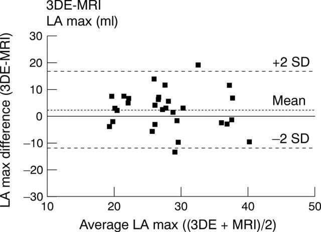
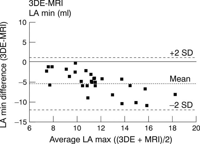
Results: Left atrial maximum and minimum volumes averaged 24.0 ml/m(2) and 7. 6 ml/m(2) by three dimensional echocardiography, and 22.1 ml/m(2) and 11.9 ml/m(2) by MRI. The greater left atrial minimum volume in the latter was at least in part a result of breath holding. Dynamic changes in left atrial volume during the heart cycle were detectable by both methods. The higher temporal resolution of three dimensional echocardiography allowed a more precise evaluation of different phases.
Conclusions: Three dimensional echocardiography and MRI were both useful methods for studying the physiological volume changes in the left atrium in children. These methods may be used for further study of the systolic and diastolic function of the heart.
Figures
Similar articles
-
Left and right atrial volume by freehand three-dimensional echocardiography: in vivo validation using magnetic resonance imaging.Eur J Echocardiogr. 2000 Mar;1(1):55-65. doi: 10.1053/euje.2000.0010. Eur J Echocardiogr. 2000. PMID: 12086217
-
Transthoracic three-dimensional echocardiography is as good as magnetic resonance imaging in measuring dynamic changes in left ventricular volume during the heart cycle in children.Eur J Echocardiogr. 2001 Mar;2(1):31-9. doi: 10.1053/euje.2000.0054. Eur J Echocardiogr. 2001. PMID: 11882423
-
Left Atrial Volumes and Function by Three-Dimensional Echocardiography: Reference Values, Accuracy, Reproducibility, and Comparison With Two-Dimensional Echocardiographic Measurements.Circ Cardiovasc Imaging. 2016 Jul;9(7):e004229. doi: 10.1161/CIRCIMAGING.115.004229. Circ Cardiovasc Imaging. 2016. PMID: 27412658
-
European Association of Cardiovascular Imaging/Cardiovascular Imaging Department of the Brazilian Society of Cardiology recommendations for the use of cardiac imaging to assess and follow patients after heart transplantation.Eur Heart J Cardiovasc Imaging. 2015 Sep;16(9):919-48. doi: 10.1093/ehjci/jev139. Epub 2015 Jul 2. Eur Heart J Cardiovasc Imaging. 2015. PMID: 26139361 Review.
-
How should we measure left atrium size and function?J Clin Ultrasound. 2012 Mar-Apr;40(3):155-66. doi: 10.1002/jcu.21871. Epub 2012 Jan 23. J Clin Ultrasound. 2012. PMID: 22271659 Review.
Cited by
-
Cardiac cycle-dependent left atrial dynamics: implications for catheter ablation of atrial fibrillation.Heart Rhythm. 2008 Jun;5(6):787-93. doi: 10.1016/j.hrthm.2008.03.003. Epub 2008 Mar 7. Heart Rhythm. 2008. PMID: 18486563 Free PMC article.
-
The Role of the Left Atrium: From Multimodality Imaging to Clinical Practice: A Review.Life (Basel). 2022 Aug 4;12(8):1191. doi: 10.3390/life12081191. Life (Basel). 2022. PMID: 36013370 Free PMC article. Review.
-
Cardiac function after repair of tetralogy of fallot: how are the atria performing? pilot study by cardiac magnetic resonance imaging.Pediatr Cardiol. 2015 Jan;36(1):96-105. doi: 10.1007/s00246-014-0970-y. Epub 2014 Aug 3. Pediatr Cardiol. 2015. PMID: 25087054
-
Normal mitral and aortic valve areas assessed by three- and two-dimensional echocardiography in 168 children and young adults.Pediatr Cardiol. 2006 Mar-Apr;27(2):217-25. doi: 10.1007/s00246-005-1056-7. Pediatr Cardiol. 2006. PMID: 16193375
-
Left atrial and left ventricular function in healthy children and young adults assessed by three dimensional echocardiography.Heart. 2003 May;89(5):544-9. doi: 10.1136/heart.89.5.544. Heart. 2003. PMID: 12695461 Free PMC article.
References
Publication types
MeSH terms
LinkOut - more resources
Full Text Sources