

Serum immunoglobulin G (IgG) and IgG subclass responses to the RgpA-Kgp proteinase-adhesin complex of Porphyromonas gingivalis in adult periodontitis
- PMID: 10768963
- PMCID: PMC97478
- DOI: 10.1128/IAI.68.5.2704-2712.2000
Serum immunoglobulin G (IgG) and IgG subclass responses to the RgpA-Kgp proteinase-adhesin complex of Porphyromonas gingivalis in adult periodontitis
Abstract
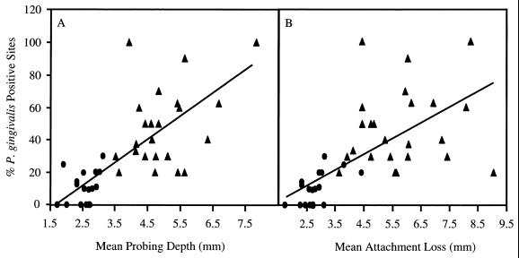
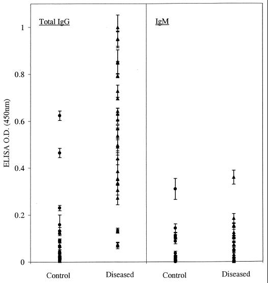
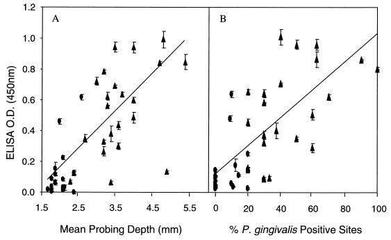
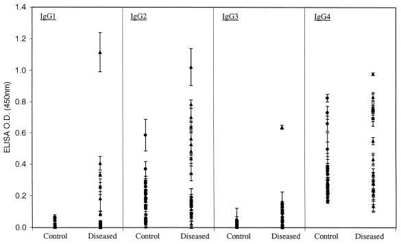
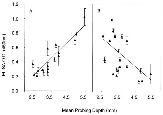
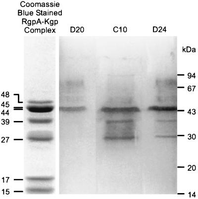
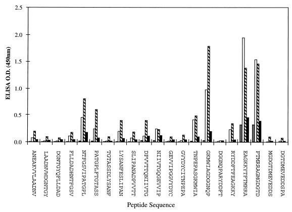
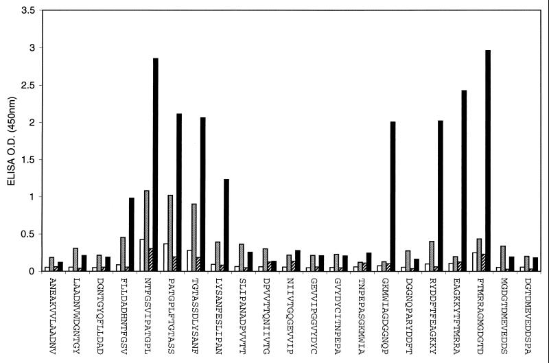
Serum immunoglobulin G (IgG), IgM, and IgG subclass responses to the RgpA-Kgp proteinase-adhesin complex of Porphyromonas gingivalis were examined by enzyme-linked immunosorbent assay using adult periodontitis patients and age- and sex-matched controls. Twenty-five sera from subjects with adult periodontitis (diseased group) and 25 sera from healthy subjects (control group) were used for the study. Sera and subgingival plaque samples from 10 sites were collected from each patient at the time of clinical examination. The level of P. gingivalis in the plaque samples was determined using a DNA probe. Highly significant positive associations between the percentage of sites positive for P. gingivalis and measures of disease severity (mean pocket depth, mean attachment loss, and percentage of sites that bled on probing) were found. The diseased group had significantly higher specific IgG responses to the RgpA-Kgp complex than did the control group, and the responses were significantly associated with mean probing depths and percentage of sites positive for P. gingivalis. Analysis of the IgG subclass responses to the RgpA-Kgp complex revealed that the subclass distribution for both the diseased and control groups was IgG4 > IgG2 > IgG3 = IgG1. The IgG2 response to the complex was positively correlated with mean probing depth, whereas the IgG4 response was negatively correlated with this measure of disease severity. Immunoblot analysis of the RgpA-Kgp complex showed that sera from healthy subjects and those with low levels of disease, with high IgG4 and low IgG2 responses, reacted with the RgpA27, Kgp39, and RgpA44 adhesins; however, sera from diseased subjects with low IgG4 and high IgG2 responses reacted only with the RgpA44 and/or Kgp44 adhesins. Epitope mapping of the RgpA27 adhesin localized a major epitope recognized by IgG4 antibodies in sera from subjects with high IgG4 and low IgG2 responses to the RgpA-Kgp complex which was not recognized by sera from diseased subjects with low IgG4 and high IgG2 responses.
Figures
 ), and IgG4 (■).
Similar articles
-
RgpA-Kgp peptide-based immunogens provide protection against Porphyromonas gingivalis challenge in a murine lesion model.Infect Immun. 2000 Jul;68(7):4055-63. doi: 10.1128/IAI.68.7.4055-4063.2000. Infect Immun. 2000. PMID: 10858222 Free PMC article.
-
Immunization with the RgpA-Kgp proteinase-adhesin complexes of Porphyromonas gingivalis protects against periodontal bone loss in the rat periodontitis model.Infect Immun. 2002 May;70(5):2480-6. doi: 10.1128/IAI.70.5.2480-2486.2002. Infect Immun. 2002. PMID: 11953385 Free PMC article.
-
Porphyromonas gingivalis RgpA-Kgp proteinase-adhesin complexes penetrate gingival tissue and induce proinflammatory cytokines or apoptosis in a concentration-dependent manner.Infect Immun. 2009 Mar;77(3):1246-61. doi: 10.1128/IAI.01038-08. Epub 2008 Dec 29. Infect Immun. 2009. PMID: 19114547 Free PMC article.
-
Porphyromonas gingivalis gingipains: the molecular teeth of a microbial vampire.Curr Protein Pept Sci. 2003 Dec;4(6):409-26. doi: 10.2174/1389203033487009. Curr Protein Pept Sci. 2003. PMID: 14683427 Review.
-
The role of gingipains in the pathogenesis of periodontal disease.J Periodontol. 2003 Jan;74(1):111-8. doi: 10.1902/jop.2003.74.1.111. J Periodontol. 2003. PMID: 12593605 Review.
Cited by
-
Unprimed, M1 and M2 Macrophages Differentially Interact with Porphyromonas gingivalis.PLoS One. 2016 Jul 6;11(7):e0158629. doi: 10.1371/journal.pone.0158629. eCollection 2016. PLoS One. 2016. PMID: 27383471 Free PMC article.
-
The leprosy reaction is associated with salivary anti-Porphyromonas gingivalis IgA antibodies.AMB Express. 2023 Jul 7;13(1):70. doi: 10.1186/s13568-023-01576-1. AMB Express. 2023. PMID: 37418096 Free PMC article.
-
GM-CSF and uPA are required for Porphyromonas gingivalis-induced alveolar bone loss in a mouse periodontitis model.Immunol Cell Biol. 2015 Sep;93(8):705-15. doi: 10.1038/icb.2015.25. Epub 2015 Mar 10. Immunol Cell Biol. 2015. PMID: 25753270
-
Humoral immune responses to S-layer-like proteins of Bacteroides forsythus.Clin Diagn Lab Immunol. 2003 May;10(3):383-7. doi: 10.1128/cdli.10.3.383-387.2003. Clin Diagn Lab Immunol. 2003. PMID: 12738635 Free PMC article.
-
Specific in situ visualization of plasma cells producing antibodies against Porphyromonas gingivalis in gingival radicular cyst: application of the enzyme-labeled antigen method.J Histochem Cytochem. 2011 Jul;59(7):673-89. doi: 10.1369/0022155411408906. Epub 2011 Apr 27. J Histochem Cytochem. 2011. PMID: 21525188 Free PMC article.
References
-
- Aalberse R C, van der Gaag R, van Leeuwen J. Serologic aspects of IgG4 antibodies. I. Prolonged immunization results in an IgG4-restricted response. J Immunol. 1983;130:722–726. - PubMed
-
- Aalberse R C, van Milligen F, Tan K Y, Stapel S O. Allergen-specific IgG4 in atopic disease. Allergy. 1993;48:559–569. - PubMed
-
- Bhogal P S, Slakeski N, Reynolds E C. A cell-associated protein complex of Porphyromonas gingivalisW50 composed of Arg- and Lys-specific cysteine proteinases and adhesins. Microbiology. 1997;143:2485–2495. - PubMed
-
- Chen Z, Potempa J, Polanowski A, Wikstrom M, Travis J. Purification and characterization of a 50-kDa cysteine proteinase (gingipain) from Porphyromonas gingivalis. J Biol Chem. 1992;267:18896–18901. - PubMed
-
- Dashper S G, O'Brien-Simpson N M, Bhogal P S, Franzmann A D, Reynolds E C. Purification and characterization of a putative fimbrial protein/receptor of Porphyromonas gingivalis. Aust Dent J. 1998;43:99–104. - PubMed
Publication types
MeSH terms
Substances
LinkOut - more resources
Full Text Sources
Other Literature Sources
