. 2000 May;68(5):2870-9.
doi: 10.1128/IAI.68.5.2870-2879.2000.

In situ analysis of the evolution of the primary immune response in murine Chlamydia trachomatis genital tract infection

Affiliations

In situ analysis of the evolution of the primary immune response in murine Chlamydia trachomatis genital tract infection

S G Morrison et al. Infect Immun. 2000 May.

Abstract

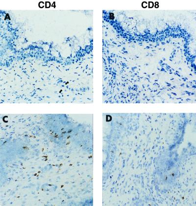
Adaptive immune responses contribute to the resolution of Chlamydia trachomatis genital tract infection and protect against reinfection, but our understanding of the mechanisms of those protective responses is incomplete. In this study, we analyzed by in situ immunohistochemistry the progression of the inflammatory and cytokine responses in the genital tracts of mice vaginally infected with C. trachomatis strain mouse pneumonitis. The cellular inflammatory response was characterized by an initial elevation in myeloid cells in the vagina (day 3) and uterine horns (day 7), followed by a marked rise in the number of T cells, predominantly CD4(+) cells. CD8(+) T cells and CD45R(+) B cells were also detected but were much less numerous. Perivascular clusters of CD4(+) T cells, which resembled clusters of T cells seen in delayed-type hypersensitivity responses, were evident by 2 weeks postinfection. Following the resolution of infection, few CD8(+) T cells and CD45R(+) B cells remained, whereas numerous CD4(+) T cells and perivascular clusters of CD4(+) T cells persisted in genital tract tissues. Interleukin-12 (IL-12)- and tumor necrosis factor alpha (TNF-alpha)-producing cells were observed in vaginal tissue by day 3 of infection and in uterine tissues by day 7. Cells producing IL-4 or IL-10 were absent from vaginal tissues at day 3 of infection but were present in uterine tissues by day 7 and were consistently more numerous than IL-12- and TNF-alpha-producing cells. Thus, the evolution of the local inflammatory response was characterized by the accumulation of CD4(+) T cells into perivascular clusters and the presence of cells secreting both Th1- and Th2-type cytokines. The persistence of CD4(+)-T-cell clusters long after infection had resolved (day 70) may provide for a readily mobilizable T-cell response by which previously infected animals can quickly respond to and control a secondary infectious challenge.


Figures

FIG. 1
FIG. 1
Time course of chlamydial genital tract infection. Mice were infected with 100 50% infectious doses of C. trachomatis strain MoPn. The course of infection was monitored by swabbing the vaginal vault at selected times and enumerating IFU by isolation onto HeLa cell monolayers. Inclusions were visualized by indirect immunofluorescence using monoclonal antibody Mo-33b and fluorescein-labeled goat anti-mouse IgG. Data are presented as mean IFU ± standard error of the mean for nine mice.
FIG. 2
FIG. 2
Immunohistochemical staining of CD11b, Ly6G, CD3e, and CD45R in vaginal tissue. Vaginal tissues were collected from uninfected mice (A to D) and from chlamydia-infected mice at 3 days post-infectious challenge (E to H) and stained with anti-CD11b (A and E), anti-Ly6G (B and F), anti-CD3e (C and G), or anti-CD45R (D and H). Magnification, ×300. Representative tissues from four or five mice at each time point are shown.
FIG. 3
FIG. 3
Immunohistochemical staining of myeloid and lymphoid cell populations in uterine tissue. At weekly intervals uterine horns were harvested from chlamydia-infected mice and stained for CD11b, Ly6G, CD3e, or CD45R cell surface antigens. (A to D) Noninfected mice; (E to H) 7 days postinfection; (I to L) 14 days postinfection; (M to P) 21 days postinfection; (Q to T and U to X) 28 days postinfection. Anti-CD11b (A, E, I, M, Q, and U), anti-Ly6G (B, F, J, N, R, and V), anti-CD3e (C, G, K, O, S, and W), and anti-CD45R (D, H, L, P, T, and X) were used. Magnifications, ×300 (A to T) and ×60 (U to X). Representative tissues from four or five mice at each time point are shown.
FIG. 4
FIG. 4
Immunohistochemical staining of CD4+- or CD8+-T-cell subsets in vaginal tissues from noninfected mice (A and B) and chlamydia-infected mice (C and D) at 3 days postinfection, stained with either anti-CD4 (A and C) or anti-CD8 (B and D). Magnification, ×300. Representative tissues from four or five mice at each time point are shown.
FIG. 5
FIG. 5
Immunohistochemical staining of either CD4+- or CD8+-T-cell subsets in uterine tissue from noninfected or chlamydia-infected mice. (A to F) Staining with anti-CD4; (G to L) staining with anti-CD8. (A and G) Noninfected; (B and H) 7 days postinfection; (C and I) 14 days postinfection; (D and J) 21 days postinfection; (E, K, F, and L) 28 days postinfection. Magnifications, ×300 (A to K) and ×60 (F and L). Representative tissues from four or five mice at each time point are shown.
FIG. 6
FIG. 6
Characterization of the local cellular inflammatory response following genital tract infection with C. trachomatis. Uterine horns were harvested at various times postinfection, processed, stained, and enumerated as described in Materials and Methods. Bars represent the mean inflammatory score ± standard deviation in uterine tissues from groups of four or five mice at each time point.
FIG. 7
FIG. 7
Immunohistochemical staining of inflammatory cells in the vagina (A to F) and uterine horns (G to L) following the resolution of chlamydial genital tract infection (day 42 postinfection). Anti-CD11b (A and G), anti-Ly6G (B and H), anti-CD45R (C and I), anti-CD3e (D and J), anti-CD4 (E and K), and anti-CD8 (F and L) were used. Magnification, ×300. Representative tissues from four or five mice are shown.
FIG. 8
FIG. 8
Immunohistochemical staining of cytokine-producing cells in the vaginal (A to H) and uterine (I to P) tissues of chlamydia-infected mice. Tissues were harvested, processed and stained as described in Materials and Methods. (A to D) Vagina, noninfected; (E to H) vagina 3 days postinfection; (I to L) uterine horn, noninfected; (M to P) uterine horn 28 days postinfection. Anti-TNF-α (A, E, I, and M), anti-IL-12 (B, F, J, and N), anti-IL-4 (C, G, K, and O), and anti-IL-10 (D, H, L, and P) were used. Representative tissues from four or five mice at each time point are shown. Magnification, ×600.

Comment in

References

    1. Barron A L, White H J, Rank R G, Soloff B L, Moses E B. A new animal model for the study of Chlamydia trachomatis genital infections: infection of mice with the agent of mouse pneumonitis. J Infect Dis. 1981;143:63–66. - PubMed
    1. Barteneva N, Theodor I, Peterson E M, de la Maza L M. Role of neutrophils in controlling early stages of a Chlamydia trachomatis infection. Infect Immun. 1996;64:4830–4833. - PMC - PubMed
    1. Cain T K, Rank R G. Local Th1-like responses are induced by intravaginal infection of mice with the mouse pneumonitis biovar of Chlamydia trachomatis. Infect Immun. 1995;63:1784–1789. - PMC - PubMed
    1. Caldwell H D, Kromhout J, Schachter J. Purification and partial characterization of the major outer membrane protein of Chlamydia trachomatis. Infect Immun. 1981;31:1161–1176. - PMC - PubMed
    1. Cotter T W, Ramsey K H, Miranpuri G S, Poulsen C E, Byrne G I. Dissemination of Chlamydia trachomatis chronic genital tract infection in gamma interferon gene knockout mice. Infect Immun. 1997;65:2145–2152. - PMC - PubMed

Publication types

MeSH terms