. 2000 Apr 17;149(2):255-62.
doi: 10.1083/jcb.149.2.255.

Cytoplasmic localization and the choice of ligand determine aggregate formation by androgen receptor with amplified polyglutamine stretch

Affiliations

Cytoplasmic localization and the choice of ligand determine aggregate formation by androgen receptor with amplified polyglutamine stretch

M Becker et al. J Cell Biol. .

Abstract

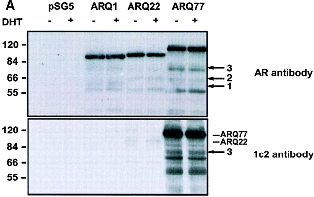
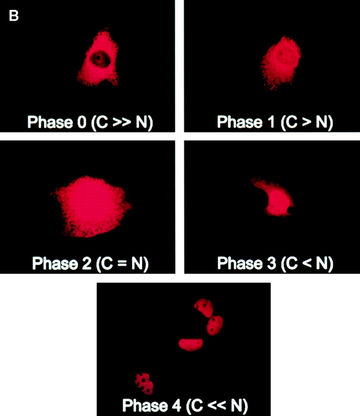
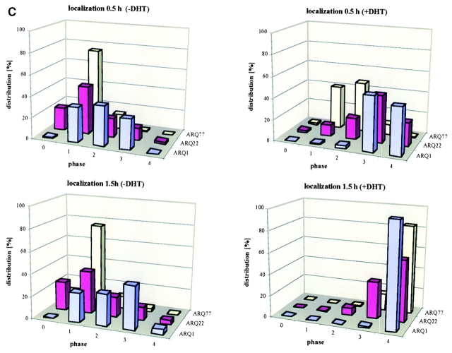
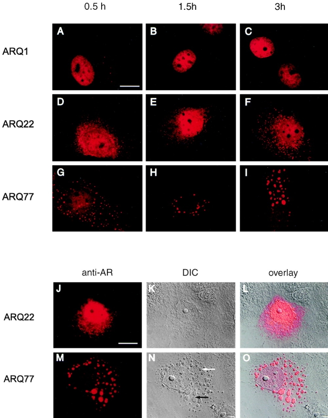
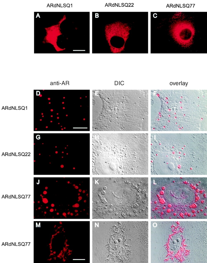
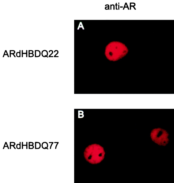
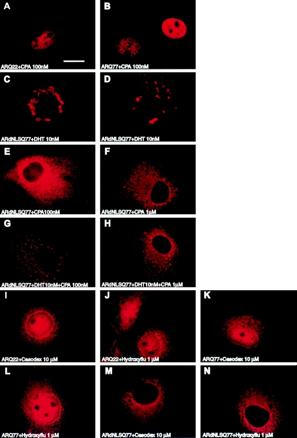
Polyglutamine tract expansion in androgen receptor is a recognized cause of spinal and bulbar muscular atrophy (SBMA), an X-linked motor neuronopathy. Similar mutations have been identified in proteins associated with other neurodegenerative diseases. Recent studies have shown that amplified polyglutamine repeat stretches form cellular aggregates that may be markers for these neurodegenerative diseases. Here we describe conditions that lead to aggregate formation by androgen receptor with polyglutamine stretch amplification. In transfection experiments, the mutant, compared with the wild-type receptor, was delayed in its cytoplasmic-nuclear translocation and formed large cytoplasmic aggregates in the presence of androgen. The cytoplasmic environment appears crucial for this aggregation, since retention of both the wild-type and mutant receptors in this cellular compartment by the deletion of their nuclear localization signals resulted in massive aggregation. Conversely, rapid nuclear transport of both receptors brought about by deletion of their ligand binding domains did not result in aggregate formation. However, androgen antagonists that altered the conformation of the ligand binding domain and promoted varying rates of cytoplasmic-nuclear translocation all inhibited aggregate formation. This demonstrates that in addition to the cytoplasmic localization, a distinct contribution of the ligand binding domain of the receptor is necessary for the aggregation. The finding that antiandrogens inhibit aggregate formation may provide the basis for in vivo determination of the role of these structures in SBMA.


Figures

Figure 1
Figure 1
Subcellular localization of ARs with polyglutamine stretch of different lengths. COS-7 cells were transfected with an empty expression vector pSG5 and AR expression vectors ARQ1, ARQ22, and ARQ77, and treated with or without DHT (10−7 M) for 0.5 or 1.5 h. (A) Western blot analysis was performed with equal amounts of cellular proteins using the anti-AR antibody AR44 and an anti-polyglutamine repeat antibody Ic2. The bands marked 1, 2, and 3 in the upper panel are NH2-terminal AR fragments as they are recognized by the AR antibody that detects an NH2-terminal epitope. In the lower panel, bands corresponding to ARQ22 and ARQ77 have been indicated. Note that the Ic2 antibody recognizes the Q77 stretch more effectively than the Q22 stretch. Also recognized by the Ic2 antibody and indicated as band 3 is a 74-kD NH2-terminal fragment of the AR containing an amplified glutamine stretch. (B) Five different phases of cellular localization of the AR defined numerically as 0–4. C, cytoplasm; N, nucleus. (C) Shown as bar diagrams are the number of cells with the transfected receptor at a particular stage of cellular localization in the presence or absence of DHT as determined by immunofluorescence microscopy.
Figure 1
Figure 1
Subcellular localization of ARs with polyglutamine stretch of different lengths. COS-7 cells were transfected with an empty expression vector pSG5 and AR expression vectors ARQ1, ARQ22, and ARQ77, and treated with or without DHT (10−7 M) for 0.5 or 1.5 h. (A) Western blot analysis was performed with equal amounts of cellular proteins using the anti-AR antibody AR44 and an anti-polyglutamine repeat antibody Ic2. The bands marked 1, 2, and 3 in the upper panel are NH2-terminal AR fragments as they are recognized by the AR antibody that detects an NH2-terminal epitope. In the lower panel, bands corresponding to ARQ22 and ARQ77 have been indicated. Note that the Ic2 antibody recognizes the Q77 stretch more effectively than the Q22 stretch. Also recognized by the Ic2 antibody and indicated as band 3 is a 74-kD NH2-terminal fragment of the AR containing an amplified glutamine stretch. (B) Five different phases of cellular localization of the AR defined numerically as 0–4. C, cytoplasm; N, nucleus. (C) Shown as bar diagrams are the number of cells with the transfected receptor at a particular stage of cellular localization in the presence or absence of DHT as determined by immunofluorescence microscopy.
Figure 2
Figure 2
Detection of cytoplasmic aggregates formed by the AR constructs ARQ1, ARQ22, and ARQ77 in COS-7 cells. COS-7 cells were transfected with ARQ1, ARQ22, and ARQ77, treated with hormones and the cellular localization of the receptors was determined by fluorescence microscopy. (A–I) Shown are some of the aggregates formed by these receptors after 0.5, 1.5, and 3 h. (J–O) A comparison of the aggregates formed by ARQ22 and ARQ77 after 3 h of DHT treatment with the use of immunoflourescence experiments (J and M), DIC (K and N), and an overlay of the two images (L and O). The white arrow points to a single small inclusion and the black arrow shows aggregates in the process of being fused. Bars, 15 μm.
Figure 3
Figure 3
Detection of cytoplasmic inclusions formed by ARdNLSQ1, ARdNLSQ22, and ARdNLSQ77 in COS-7 cells. COS-7 cells were transfected with the indicated receptor constructs lacking the NLS, treated with (D–O) or without (A–C) DHT, and the cellular localization of the receptors assessed by fluorescence microscopy. Shown are the different types of aggregates formed as detected by immunofluorescence microscopy, DIC images, and an overlay of the two pictures. Bars: 15 μm (A–C); 30 μm (D–L); and 15 μm (M–O).
Figure 4
Figure 4
Androgen receptor lacking the LBD does not form aggregates. COS-7 cells were transfected with ARQ22 and ARQ77 lacking the LBD (ARdHBDQ22, ARdHBDQ77) and cellular localization of the receptor was determined by fluorescence microscopy.
Figure 5
Figure 5
Cytoplasmic aggregation of the AR is not induced by antiandrogens. COS-7 cells were transfected with the receptor constructs ARQ22, ARQ77, and their counterparts lacking the NLS (ARdNLSQ22, ARdNLSQ77). The transfected cells were treated with the indicated concentrations of DHT and the antiandrogens cyproterone acetate (CPA), hydroxyflutamide (Hydroxyflu) or casodex for 3 h. The effect of these ligands on aggregate formation was detected by immunofluorescence microscopy. Bar, 15 μm.

Similar articles

Cited by

References

    1. Bingham P.M., Scott M.O., Wang S., McPhaul M.J., Wilson E.M., Garbern J.Y., Merry D.E., Fischbeck K.H. Stability of an expanded trinucleotide repeat in the androgen receptor gene in transgenic mice. Nat. Genet. 1995;9:191–196. - PubMed
    1. Butler R., Leigh P.N., McPhaul M.J., Gallo J.-M. Truncated forms of the androgen receptor are associated with polyglutamine expansion in X-linked spinal and bulbar muscular atrophy. Hum. Mol. Genet. 1998;7:121–127. - PubMed
    1. Chamberlain N.L., Driver E.D., Miesfeld R.L. The length and location of CAG trinucleotide repeats in the androgen receptor N-terminal domain affect transactivation function. Nucleic Acids Res. 1994;22:3181–3186. - PMC - PubMed
    1. Culig Z., Hobisch A., Cronauer M.V., Cato A.C.B., Hitmair A., Radmayr C., Eberle J., Bartsch G., Klocker H. Mutant androgen receptor detected in an advanced-stage prostatic carcinoma is activated by adrenal androgens and progesterone. Mol. Endocrinol. 1993;7:1541–1550. - PubMed
    1. Danek A., Witt T.N., Mann K., Schweikert H.U., Romalo G., La Spada A.R., Fischbeck K.H. Decrease in androgen binding and effect of androgen treatment in a case of X-linked bulbospinal neuronopathy. Clin. Investig. 1994;72:892–897. - PubMed

Publication types