. 2000 Apr 17;149(2):423-30.
doi: 10.1083/jcb.149.2.423.

Suppression of Pyk2 kinase and cellular activities by FIP200

Affiliations

Suppression of Pyk2 kinase and cellular activities by FIP200

H Ueda et al. J Cell Biol. .

Abstract

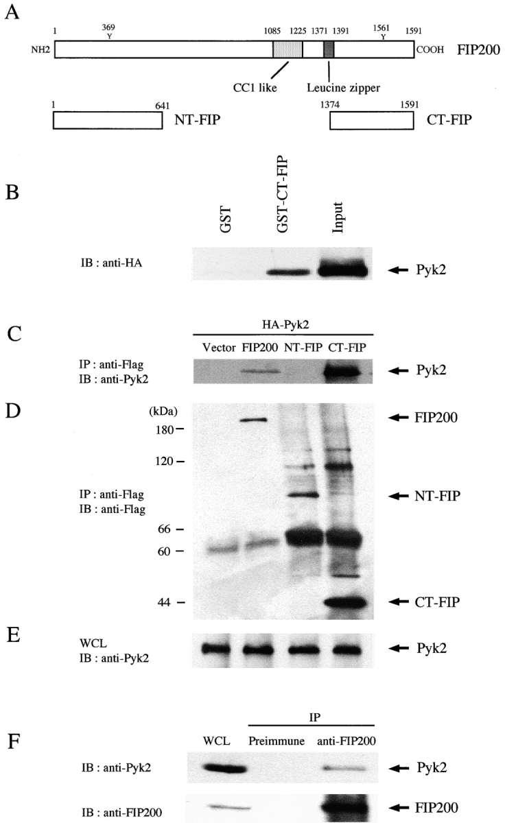
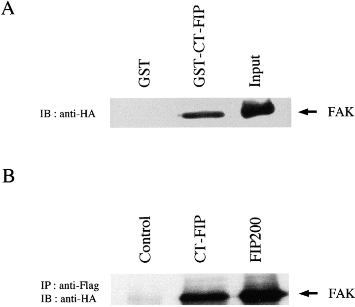
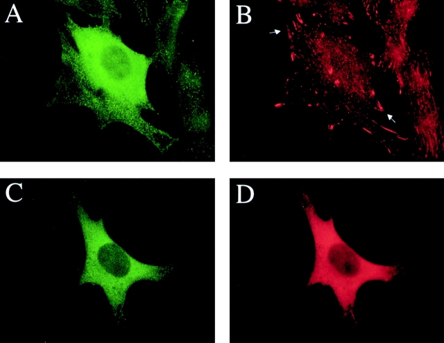
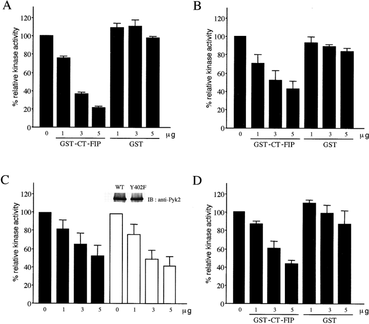
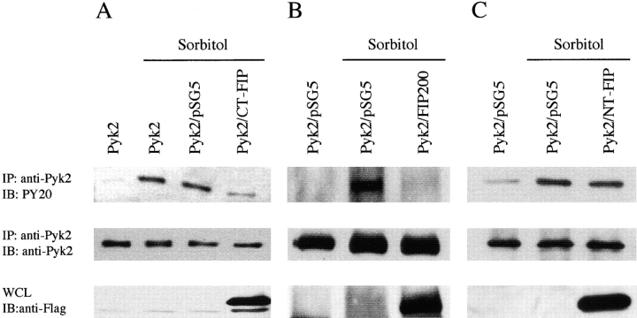
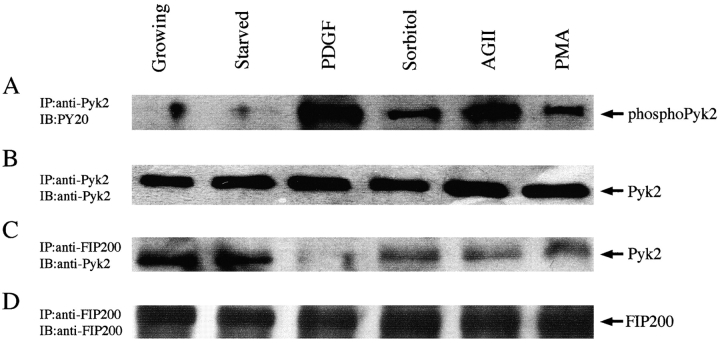
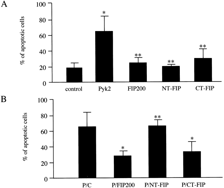
Proline-rich tyrosine kinase 2 (Pyk2) is a cytoplasmic tyrosine kinase implicated to play a role in several intracellular signaling pathways. We report the identification of a novel Pyk2-interacting protein designated FIP200 (FAK family kinase-interacting protein of 200 kD) by using a yeast two-hybrid screen. In vitro binding assays and coimmunoprecipitation confirmed association of FIP200 with Pyk2, and similar assays also showed FIP200 binding to FAK. However, immunofluorescent staining indicated that FIP200 was predominantly localized in the cytoplasm. FIP200 bound to the kinase domain of Pyk2 and inhibited its kinase activity in in vitro kinase assays. FIP200 also inhibited the kinase activity of the Pyk2 isolated from SYF cells (deficient in Src, Yes, and Fyn expression) and the Pyk2 mutant lacking binding site for Src, suggesting that it regulated Pyk2 kinase directly rather than affecting the associated Src family kinases. Consistent with its inhibitory effect in vitro, FIP200 inhibited activation of Pyk2 and Pyk2-induced apoptosis in intact cells, which correlated with its binding to Pyk2. Finally, activation of Pyk2 by several biological stimuli correlated with the dissociation of endogenous FIP200-Pyk2 complex, which provided further support for inhibition of Pyk2 by FIP200 in intact cells. Together, these results suggest that FIP200 functions as an inhibitor of Pyk2 via binding to its kinase domain.


Figures

Figure 1
Figure 1
Association of Pyk2 with FIP200 COOH-terminal fragment. (A) A schematic of FIP200 is shown on top. The NH2- and COOH-terminal FIP200 are shown below. (B) Immobilized GST-CT-FIP or GST was incubated with lysates from 293T cells that had been transfected with pKH3-Pyk2. After washing, the bound proteins were analyzed by Western blotting with anti-HA. An aliquot of the lysate was also analyzed directly (input). (C–E) 293T cells were cotransfected with pKH3-Pyk2 and pSG5 vectors encoding Flag-tagged FIP200, NT-FIP, CT-FIP, or control vector, as indicated. 2 d after transfection, the cell lysates were immunoprecipitated by anti-Flag and analyzed by Western blotting with either anti-Pyk2 (C) or anti-Flag (D). Aliquots of the lysates were also analyzed directly by Western blotting with anti-Pyk2 (E). (F) Lysates from RASM cells were immunoprecipitated by anti-FIP200 or preimmune serum as indicated. The immune complexes or an aliquot of the lysate (WCL) were analyzed by Western blotting with anti-Pyk2 (top panel) or anti-FIP200 (bottom panel). Molecular mass markers are shown on the left, and the positions of Pyk2, FIP200, and its fragments are marked on the right.
Figure 2
Figure 2
FIP200 association with FAK. (A) Immobilized GST-CT-FIP or GST alone was incubated with lysates from 293T cells that had been transfected with pKH3-FAK. After washing, the bound proteins were analyzed by Western blotting with anti-HA. An aliquot of the lysate was also analyzed directly by Western blotting with anti-HA (input). (B) 293T cells were cotransfected with pKH3-FAK and pSG5 vectors encoding Flag-tagged FIP200, CT-FIP, or pSG5 vector alone (control), as indicated. The lysates were immunoprecipitated with anti-Flag and analyzed by Western blotting with anti-HA. The positions of transfected FAK are marked by arrows.
Figure 3
Figure 3
Subcellular localization of FIP200. NIH3T3 cells were transfected with vector encoding Flag-FIP200 (A and B) or both vectors encoding Flag-FIP200 and HA-Pyk2 (C and D). 1 d after transfection, cells were processed for immunofluorescent staining as described in Materials and Methods. The primary antibodies used are anti-Flag (A and C), antivinculin (B), or anti-HA (D). Examples of focal contacts are marked by arrows in B.
Figure 4
Figure 4
Inhibition of Pyk2 kinase activity by CT-FIP. (A) Pyk2 was immunoprecipitated from RASM cell lysates. Aliquots of the immune complex were assayed for kinase activity using E4Y1 as an exogenous substrate in the presence of various amounts of GST-CT-FIP or GST as indicated. The region containing the labeled E4Y1 was subjected to quantitation using a PhosphoImager as described in Materials and Methods. Relative kinase activities were normalized to Pyk2 activity in the absence of the GST fusion protein. The mean and SD of the relative kinase activities from three independent experiments are shown. (B) The effect of GST-CT-FIP or GST alone on the kinase activity of Pyk2 immunoprecipitated from SYF cells was examined as described in A. The mean and SD of the relative kinase activities from three independent experiments are shown. (C) 293T cells were transfected with pKH3-Pyk2 (filled bars) or pKH3-Pyk2Y402F (open bars). They were immunoprecipitated and assayed for kinase activities in the presence of various amounts of GST-CT-FIP as described in A. The mean and SD of the relative kinase activities from three independent experiments are shown. The inset shows comparable expression levels of the transfected Pyk2 and Y402F mutant. (D) Aliquots of the FAK immunoprecipitates from NIH3T3 cells were assayed for kinase activity using E4Y1 as an exogenous substrate in the presence of various amounts of GST-CT-FIP or GST as indicated. The activity was quantified as described in A.
Figure 5
Figure 5
FIP200 inhibition of Pyk2 tyrosine phosphorylation in response to sorbitol. CHO cells were cotransfected with pKH3-Pyk2 and pSG5 vectors encoding FIP200, NT-FIP, CT-FIP, or pSG5 alone, as indicated. 2 d after transfection, cells were serum-starved for 24 h and treated with sorbitol (400 mM for 5 min). Lysates were immunoprecipitated by anti-Pyk2 and analyzed by Western blotting with either PY20 to assay the phosphorylation (top) or anti-Pyk2 to verify the expression levels of Pyk2 (middle). Aliquots of the lysates were also analyzed directly by Western blotting with anti-Flag to detect FIP200 or its fragments (bottom).
Figure 6
Figure 6
Pyk2 phosphorylation and its association with FIP200 in response to different stimuli. Serum-starved RASM cells were stimulated with various agents as indicated (PDGF, 25 ng/ml for 5 min; sorbitol, 400 mM for 10 min; AGII, 1 μM angiotensin II for 1.5 min; and PMA, 200 nM for 5 min). Cell lysates were collected and immunoprecipitated with either anti-Pyk2 (A and B) or anti-FIP200 (C and D) antibodies. The immune complexes were Western-blotted with PY20 to assay for Pyk2 phosphorylation (A) and anti-Pyk2 to detect FIP200–Pyk2 association (C), or anti-Pyk2 (B) and anti-FIP200 (D) to verify similar immunoprecipitation in all samples.
Figure 7
Figure 7
FIP200 inhibition of Pyk2-induced apoptosis. (A) Rat-1 cells were cotransfected with a plasmid encoding GFP and expression vectors encoding Pyk2, FIP200, NT-FIP, CT-FIP, or vector alone (control), as indicated. The results show the mean and SD of a percentage of apoptotic cells among the positively transfected (GFP+) cells from at least four independent experiments. *P < 0.01 and **P ≥ 0.15 in comparison to the value from cells transfected with the control vector (Minitab Release 10.5 Xtra). (B) Rat-1 cells were cotransfected with a plasmid encoding GFP, pKH3-Pyk2 (P), and expression vectors encoding FIP200, NT-FIP, CT-FIP, or vector alone (C), as indicated. They were analyzed as in A, and the results show the mean and SD of a percentage of apoptotic/GFP positive cells. *P < 0.01 and **P > 0.40 in comparison to the value from cells transfected with Pyk2 and a control vector (Minitab Release 10.5 Xtra).

References

    1. Andreev J., Simon J.P., Sabatini D.D., Kam J., Plowman G., Randazzo P.A., Schlessinger J. Identification of a new Pyk2 target protein with Arf-GAP activity. Mol. Cell. Biol. 1999;19:2338–2350. - PMC - PubMed
    1. Astier A., Avraham H., Manie S.N., Groopman J., Canty T., Avraham S., Freedman A.S. The related adhesion focal tyrosine kinase is tyrosine-phosphorylated after beta1-integrin stimulation in B cells and binds to p130cas. J. Biol. Chem. 1997;272:228–232. - PubMed
    1. Avraham S., London R., Fu Y., Ota S., Hiregowdara D., Li J., Jiang S., Pasztor L.M., White R.A., Groopman J.E. Identification and characterization of a novel related adhesion focal tyrosine kinase (RAFTK) from megakaryocytes and brain. J. Biol. Chem. 1995;270:27742–27751. - PubMed
    1. Brinson A.E., Harding T., Diliberto P.A., He Y., Li X., Hunter D., Herman B., Earp H.S., Graves L.M. Regulation of a calcium-dependent tyrosine kinase in vascular smooth muscle cells by angiotensin II and platelet-derived growth factor. Dependence on calcium and the actin cytoskeleton. J. Biol. Chem. 1998;273:1711–1718. - PubMed
    1. Cary L.A., Chang J.F., Guan J.L. Stimulation of cell migration by overexpression of focal adhesion kinase and its association with Src and Fyn. J. Cell Sci. 1996;109:1787–1794. - PubMed

Publication types

MeSH terms