

Identification and expression of genes involved in biosynthesis of L-oleandrose and its intermediate L-olivose in the oleandomycin producer Streptomyces antibioticus
- PMID: 10770761
- PMCID: PMC89854
- DOI: 10.1128/AAC.44.5.1266-1275.2000
Identification and expression of genes involved in biosynthesis of L-oleandrose and its intermediate L-olivose in the oleandomycin producer Streptomyces antibioticus
Abstract
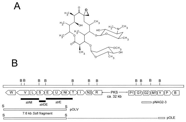
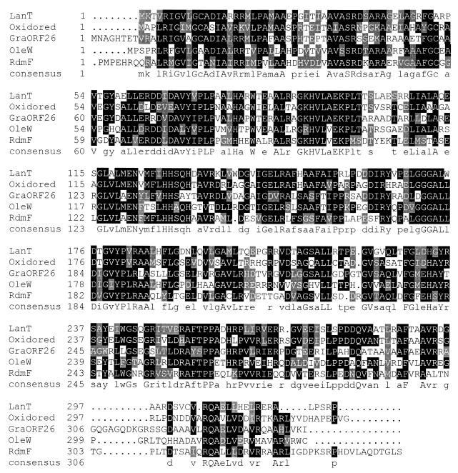
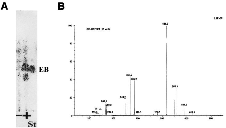
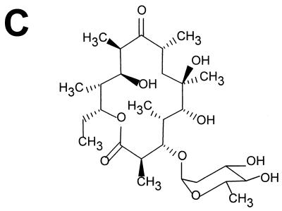
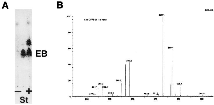
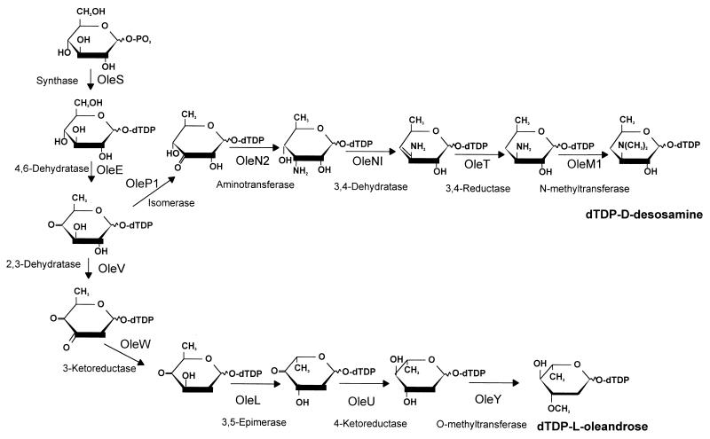
A 9.8-kb DNA region from the oleandomycin gene cluster in Streptomyces antibioticus was cloned. Sequence analysis revealed the presence of 8 open reading frames encoding different enzyme activities involved in the biosynthesis of one of the two 2, 6-deoxysugars attached to the oleandomycin aglycone: L-oleandrose (the oleW, oleV, oleL, and oleU genes) and D-desosamine (the oleNI and oleT genes), or of both (the oleS and oleE genes). A Streptomyces albus strain harboring the oleG2 glycosyltransferase gene integrated into the chromosome was constructed. This strain was transformed with two different plasmid constructs (pOLV and pOLE) containing a set of genes proposed to be required for the biosynthesis of dTDP-L-olivose and dTDP-L-oleandrose, respectively. Incubation of these recombinant strains with the erythromycin aglycon (erythronolide B) gave rise to two new glycosylated compounds, identified as L-3-O-olivosyl- and L-3-O-oleandrosyl-erythronolide B, indicating that pOLV and pOLE encode all enzyme activities required for the biosynthesis of these two 2,6-dideoxysugars. A pathway is proposed for the biosynthesis of these two deoxysugars in S. antibioticus.
Figures

References
-
- Altschul S F, Gish W, Miller W, Myers E W, Lipman D J. Basic local alignment search tool. J Mol Biol. 1990;215:403–410. - PubMed
-
- August P R, Tang L, Yoon Y J, Ning S, Muller R, Yu T W, Taylor M, Hoffmann D, Kim G G, Zhang X, Hutchinson C R, Floss H G. Biosynthesis of the ansamycin antibiotic rifamycin: deductions from the molecular analysis of the rif biosynthetic gene cluster of Amycolatopsis mediterraneiS699. Chem Biol. 1998;5:69–79. - PubMed
-
- Bauer A J, Rayment I, Frey P A, Holden H M. The molecular structure of UDP-galactose-4-epimerase from Escherichia colidetermined at 2.5 Å resolution. Proteins. 1992;12:372–381. - PubMed
-
- Bechthold A, Sohng J K, Smith T M, Chu X, Floss H G. Identification of Streptomyces violaceoruber Tü22 genes involved in the biosynthesis of granaticin. Mol Gen Genet. 1995;248:610–620. - PubMed
-
- Bullock W O, Fernández J M, Short J M. XL1-Blue: a high efficiency plasmid transforming recA Escherichia colistrain with β-galactosidase selection. BioTechniques. 1987;5:376.
Publication types
MeSH terms
Substances
Associated data
- Actions
LinkOut - more resources
Full Text Sources
Other Literature Sources
Molecular Biology Databases
