. 2000 Apr 17;191(8):1381-94.
doi: 10.1084/jem.191.8.1381.

Localization of distinct Peyer's patch dendritic cell subsets and their recruitment by chemokines macrophage inflammatory protein (MIP)-3alpha, MIP-3beta, and secondary lymphoid organ chemokine

Affiliations

Localization of distinct Peyer's patch dendritic cell subsets and their recruitment by chemokines macrophage inflammatory protein (MIP)-3alpha, MIP-3beta, and secondary lymphoid organ chemokine

A Iwasaki et al. J Exp Med. .

Abstract

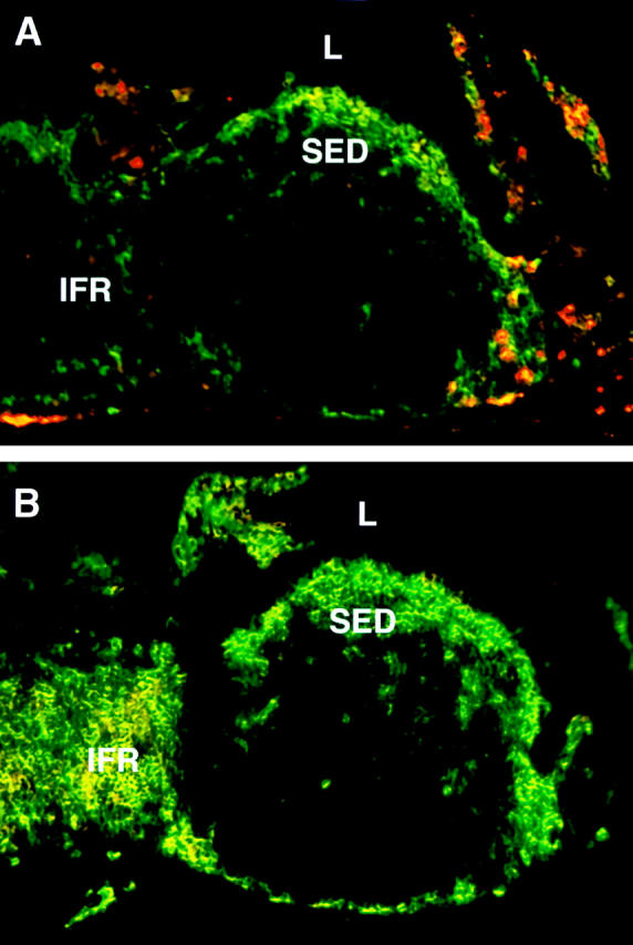
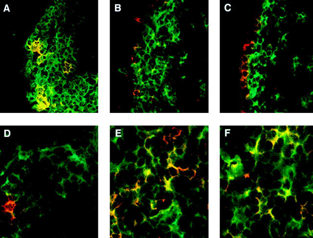
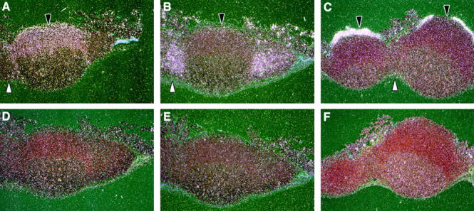
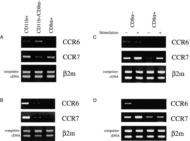
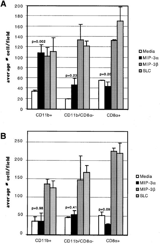
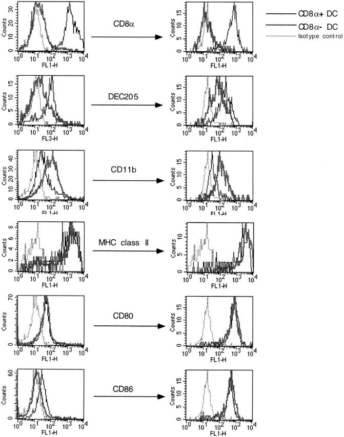
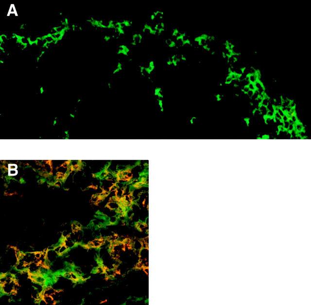
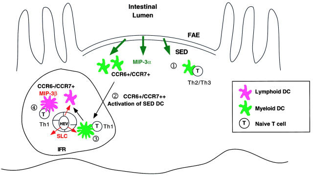
We describe the anatomical localization of three distinct dendritic cell (DC) subsets in the murine Peyer's patch (PP) and explore the role of chemokines in their recruitment. By two-color in situ immunofluorescence, CD11b(+) myeloid DCs were determined to be present in the subepithelial dome (SED) region, whereas CD8alpha(+) lymphoid DCs are present in the T cell-rich interfollicular region (IFR). DCs that lack expression of CD8alpha or CD11b (double negative) are present in both the SED and IFR. By in situ hybridization, macrophage inflammatory protein (MIP)-3alpha mRNA was dramatically expressed only by the follicle-associated epithelium overlying the SED, while its receptor, CCR6, was concentrated in the SED. In contrast, CCR7 was expressed predominantly in the IFR. Consistent with these findings, reverse transcriptase polymerase chain reaction analysis and in vitro chemotaxis assays using freshly isolated DCs revealed that CCR6 was functionally expressed only by DC subsets present in the SED, while all subsets expressed functional CCR7. Moreover, none of the splenic DC subsets migrated toward MIP-3alpha. These data support a distinct role for MIP-3alpha/CCR6 in recruitment of CD11b(+) DCs toward the mucosal surfaces and for MIP-3beta/CCR7 in attraction of CD8alpha(+) DCs to the T cell regions. Finally, we demonstrated that all DC subsets expressed an immature phenotype when freshly isolated and maintained expression of subset markers upon maturation in vitro. In contrast, CCR7 expression by myeloid PP DCs was enhanced with maturation in vitro. In addition, this subset disappeared from the SED and appeared in the IFR after microbial stimulation in vivo, suggesting that immature myeloid SED DCs capture antigens and migrate to IFR to initiate T cell responses after mucosal microbial infections.


Figures

Figure 1
Figure 1
Localization of myeloid and lymphoid DCs in the PP. Frozen sections of PPs were doubly stained with antibodies against CD11c (green) in combination with (A) anti-CD11b (red) or (B) anti-CD8α (red). L, lumen.
Figure 2
Figure 2
Confocal microscopic analysis of myeloid and lymphoid DCs in the PP. Cryostat sections of mouse PPs were doubly labeled with antibodies against CD11c (green) in combination with (A and D) anti-CD11b (red); (B and E) anti-CD8α (red); or (C and F) anti–DEC-205. SED regions (A–C) and IFRs (D–F) were analyzed by confocal microscopy.
Figure 3
Figure 3
Expression of MIP-3α, CCR6, and CCR7 in the PP. Antisense and sense riboprobes were hybridized to paraffin-embedded sections of the PP. The radioactive RNA probe bound to tissue section was detected by emulsion autoradiography. Darkfield images are shown for antisense CCR6 (A), CCR7 (B), and MIP-3α (C) and sense CCR6 (D), CCR7 (E), and MIP-3α (F) in the PP. Black arrows indicate the SED, and white arrows point toward IFR.
Figure 4
Figure 4
Semiquantitative RT-PCR for CCR6 and CCR7 mRNA on FACS®-sorted fresh and stimulated DC subsets from the PP and spleen. Total RNA was isolated from FACS®-sorted CD11b+, CD8α+, or DN PP (A) or spleen (B) freshly isolated DCs. RNA was also isolated from either freshly isolated or in vitro–stimulated CD8α+ or CD8α PP (C) or spleen (D) DCs. The cDNA was prepared, and the amount used for each PCR was equalized by competitive PCR using primers for the control gene (β2m) (bottom panels). The levels of CCR6 (top panels) and CCR7 (middle panels) mRNA expression were determined using specific primers as described in Materials and Methods. The amplification fragments were visualized on 1% agarose gel using ethidium bromide. Similar results were obtained from three independent experiments.
Figure 5
Figure 5
Chemotactic capacity of DC subsets toward MIP-3α and MIP-3β. FACS®-purified myeloid (CD11b+), DN (CD11b/CD8α2), and lymphoid (CD8α1) DCs from PP (A) or spleen (B) were placed in the chemotaxis chamber in duplicates. Results are expressed as the average number of cells migrating to the bottom chamber per field. At least five independent fields were counted per bottom chamber. The P values for DC migration to MIP-3α compared with the media control are indicated. The data is representative of five independent experiments.
Figure 6
Figure 6
Flow cytometric analysis of CD8α+ and CD8α PP DCs upon stimulation in vitro. Highly purified CD8α+ and CD8α DCs from PP were isolated using flow cytometric sorting, and surface expression markers on freshly isolated (left) and in vitro–activated (right) DCs were determined by flow cytometry. The same concentrations of antibodies were used to stain the DCs at left and right. This figure is a representative of three similar experiments.
Figure 7
Figure 7
In vivo migration of myeloid DCs from the SED to the IFR of the PP upon microbial stimulation. Cryostat sections of PPs were obtained from mice injected systemically with STAg 6 h earlier and were doubly labeled with antibodies against CD11c (green) in combination with anti-CD11b (red). The SED region (A) and IFR (B) were analyzed by confocal microscopy. The experiment was repeated twice with similar data.
Figure 8
Figure 8
Proposed mechanism of DC recruitment to the distinct regions of the PP. The CD11b+ DC (CCR6+/CCR7+) and CD8α+ DC (CCR6/CCR7+) precursors from the blood enter the PP by responding to CCR7 ligands secreted by the IFR. The myeloid DCs acquire responsiveness to MIP-3α due to the high levels of TGF-β present in the PPs and are thus recruited toward the FAE secreting MIP-3α. On the other hand, lymphoid DCs remain in the IFR due to their expression of CCR7 but not CCR6. Upon encountering luminal antigen transported via M cells, SED DCs may undergo two distinct differentiation pathways. If the antigen encountered is an innocuous food protein, the default pathway for CD11b+ DCs is to generate Th2/Th3 responses through secretion of high levels of IL-10 and TGF-β and low levels of IL-12 in the SED ○1. However, upon encounter with microbial stimuli, such as double-stranded RNA or LPS (“danger signals”), conventional maturation of DCs is triggered ○2. This maturation of SED DCs leads to their migration toward the IFR by upregulating CCR7 expression. In the IFR, naive T cells are primed to secrete IFN-γ by antigen presented by the CD11b+ DCs directly ○3. Alternatively, antigens processed by CD11b+ DCs may be transferred to the Th1-inducing CD8α+ DCs for subsequent presentation to naive T cells by a process known as “cross-priming” ○4.

References

    1. Spalding D.M., Griffin J.A. Different pathways of differentiation of pre-B cell lines are induced by dendritic cells and T cells from different lymphoid tissues. Cell. 1986;44:507–515. - PubMed
    1. Kelsall B.L., Strober W. Distinct populations of dendritic cells are present in the subepithelial dome and T cell regions of the murine Peyer's patch. J. Exp. Med. 1996;183:237–247. - PMC - PubMed
    1. Iwasaki A., Kelsall B.L. Freshly isolated Peyer's patch, but not spleen, dendritic cells produce interleukin 10 and induce the differentiation of T helper type 2 cells. J. Exp. Med. 1999;190:229–240. - PMC - PubMed
    1. Ardavin C., Wu L., Li C.L., Shortman K. Thymic dendritic cells and T cells develop simultaneously in the thymus from a common precursor population. Nature. 1993;362:761–763. - PubMed
    1. Suss G., Shortman K. A subclass of dendritic cells kills CD4 T cells via Fas/Fas-ligand-induced apoptosis. J. Exp. Med. 1996;183:1789–1796. - PMC - PubMed

Publication types

MeSH terms