

Entry of B cell receptor into signaling domains is inhibited in tolerant B cells
- PMID: 10770810
- PMCID: PMC2193133
- DOI: 10.1084/jem.191.8.1443
Entry of B cell receptor into signaling domains is inhibited in tolerant B cells
Abstract
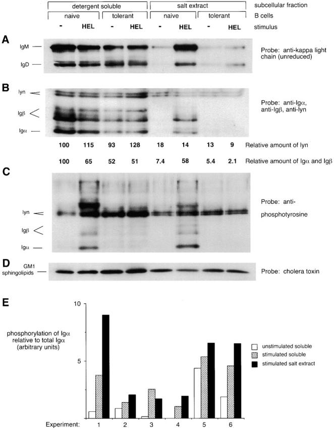
Signal transduction through the B cell antigen receptor (BCR) is altered in B cells that express a receptor that recognizes self-antigen. To understand the molecular basis for the change in signaling in autoreactive B cells, a transgenic model was used to isolate a homogeneous population of tolerant B lymphocytes. These cells were compared with a similar population of naive B lymphocytes. We show that the BCR from naive B cells enters a detergent-insoluble domain of the cell within 6 s after antigen binding, before a detectable increase in BCR phosphorylation. This fraction appears to be important for signaling because it is enriched for lyn kinase but lacks CD45 tyrosine phosphatase and because the BCR that moves into this domain becomes more highly phosphorylated. Partitioning of the BCR into this fraction is unaffected by src family kinase inhibition. Tolerant B cells do not efficiently partition the BCR into the detergent-insoluble domain, providing an explanation for their reduced tyrosine kinase activation and calcium flux in response to antigen. These results identify an early, regulated step in antigen receptor signaling and self-tolerance.
Figures


Similar articles
-
Functional analysis of Csk in signal transduction through the B-cell antigen receptor.Mol Cell Biol. 1994 Nov;14(11):7306-13. doi: 10.1128/mcb.14.11.7306-7313.1994. Mol Cell Biol. 1994. PMID: 7935444 Free PMC article.
-
Polygenic autoimmune traits: Lyn, CD22, and SHP-1 are limiting elements of a biochemical pathway regulating BCR signaling and selection.Immunity. 1998 Apr;8(4):497-508. doi: 10.1016/s1074-7613(00)80554-3. Immunity. 1998. PMID: 9586639
-
Defective negative regulation of antigen receptor signaling in Lyn-deficient B lymphocytes.Curr Biol. 1998 May 7;8(10):545-53. doi: 10.1016/s0960-9822(98)70223-4. Curr Biol. 1998. PMID: 9601638
-
The Alternate Pathway for BCR Signaling Induced by IL-4 Requires Lyn Tyrosine Kinase.J Mol Biol. 2021 Jan 8;433(1):166667. doi: 10.1016/j.jmb.2020.10.002. Epub 2020 Oct 13. J Mol Biol. 2021. PMID: 33058880 Review.
-
Signal transduction by the B-cell antigen receptor.Ann N Y Acad Sci. 1995 Sep 7;766:195-201. doi: 10.1111/j.1749-6632.1995.tb26662.x. Ann N Y Acad Sci. 1995. PMID: 7486656 Review.
Cited by
-
Antigenic liposomes displaying CD22 ligands induce antigen-specific B cell apoptosis.J Clin Invest. 2013 Jul;123(7):3074-83. doi: 10.1172/JCI69187. Epub 2013 Jun 3. J Clin Invest. 2013. PMID: 23722906 Free PMC article.
-
Transmodulation of BCR signaling by transduction-incompetent antigen receptors: implications for impaired signaling in anergic B cells.J Immunol. 2002 May 1;168(9):4344-51. doi: 10.4049/jimmunol.168.9.4344. J Immunol. 2002. PMID: 11970976 Free PMC article.
-
Functional anergy in a subpopulation of naive B cells from healthy humans that express autoreactive immunoglobulin receptors.J Exp Med. 2009 Jan 16;206(1):139-51. doi: 10.1084/jem.20080611. Epub 2008 Dec 22. J Exp Med. 2009. PMID: 19103878 Free PMC article.
-
Involvement of LAT, Gads, and Grb2 in compartmentation of SLP-76 to the plasma membrane.J Exp Med. 2000 Sep 18;192(6):847-56. doi: 10.1084/jem.192.6.847. J Exp Med. 2000. PMID: 10993915 Free PMC article.
-
Ligand-dependent and -independent processes in B-cell-receptor-mediated signaling.Springer Semin Immunopathol. 2001 Dec;23(4):333-50. doi: 10.1007/s281-001-8163-6. Springer Semin Immunopathol. 2001. PMID: 11826613 Review.
References
-
- Goodnow C.C., Crosbie J., Adelstein S., Lavoie T.B., Smith-Gill S.J., Brink R.A., Pritchard-Briscoe H., Wotherspoon J.S., Loblay R.H., Raphael K. Altered immunoglobulin expression and functional silencing of self-reactive B lymphocytes in transgenic mice. Nature. 1988;334:676–682. - PubMed
-
- Goodnow C.C., Brink R., Adams E. Breakdown of self-tolerance in anergic B lymphocytes. Nature. 1991;352:532–536. - PubMed
-
- Cyster J.G., Hartley S.B., Goodnow C.C. Competition for follicular niches excludes self-reactive cells from the recirculating B-cell repertoire. Nature. 1994;371:389–395. - PubMed
-
- Healy J.I., Dolmetsch R.E., Timmerman L.A., Cyster J.G., Thomas M.L., Crabtree G.R., Lewis R.S., Goodnow C.C. Different nuclear signals are activated by the B cell receptor during positive versus negative signaling. Immunity. 1997;6:419–428. - PubMed
Publication types
MeSH terms
Substances
Grants and funding
LinkOut - more resources
Full Text Sources
Research Materials
Miscellaneous
