. 2000 Apr;105(8):1049-56.
doi: 10.1172/JCI9259.

Targeted disruption of the class B scavenger receptor CD36 protects against atherosclerotic lesion development in mice

Affiliations

Targeted disruption of the class B scavenger receptor CD36 protects against atherosclerotic lesion development in mice

M Febbraio et al. J Clin Invest. 2000 Apr.

Abstract

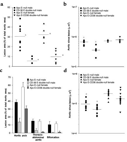
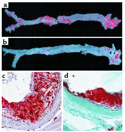
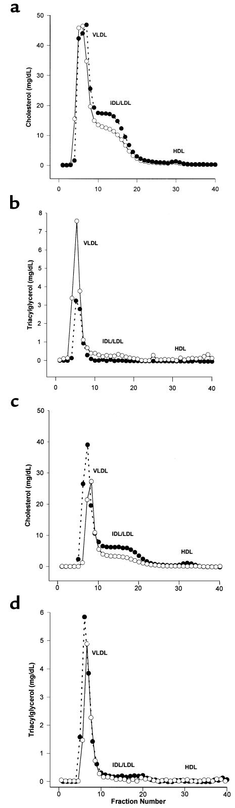
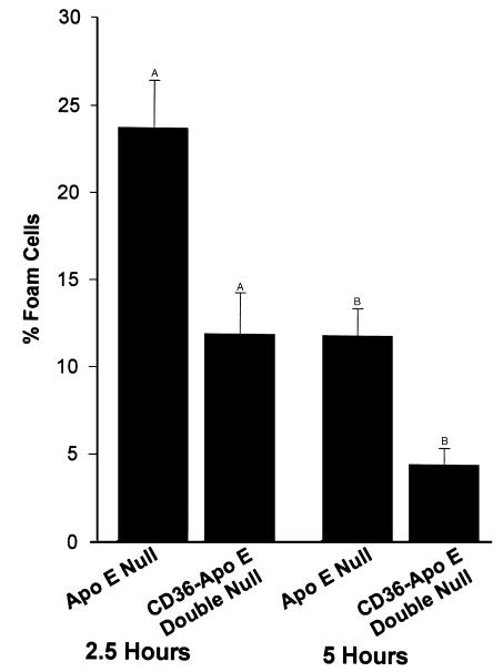
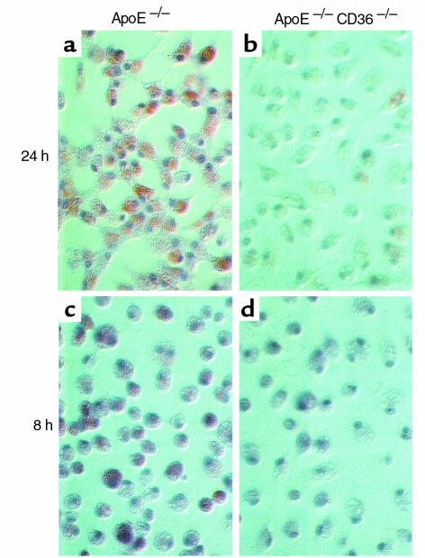
Macrophage scavenger receptors have been implicated as key players in the pathogenesis of atherosclerosis. To assess the role of the class B scavenger receptor CD36 in atherogenesis, we crossed a CD36-null strain with the atherogenic apo E-null strain and quantified lesion development. There was a 76.5% decrease in aortic tree lesion area (Western diet) and a 45% decrease in aortic sinus lesion area (normal chow) in the CD36-apo E double-null mice when compared with controls, despite alterations in lipoprotein profiles that often correlate with increased atherogenicity. Macrophages derived from CD36-apo E double-null mice bound and internalized more than 60% less copper-oxidized LDL and LDL modified by monocyte-generated reactive nitrogen species. A similar inhibition of in vitro lipid accumulation and foam cell formation after exposure to these ligands was seen. These results support a major role for CD36 in atherosclerotic lesion development in vivo and suggest that blockade of CD36 can be protective even in more extreme proatherogenic circumstances.


Figures

Figure 1
Figure 1
Disruption of CD36 decreases atherosclerotic lesion development in apo E–null mice. Circles, apo E-null male (n = 9); squares, CD36-apo E double-null male (n = 5); triangle, apo E–null female (n = 5); reversed triangle, CD36-apo E double-null female (n = 6). (a) Lesion surface area in the aortic tree was assessed by en face oil red-O positive staining. The aortic tree from the heart to below the bifurcation of the iliac arteries was removed and dissected for staining and lesion analysis from mice fed a Western diet for 12 weeks. Each sample was scanned, and total oil red-O positive area was determined using Scion Image software. AP < 0.001, BP < 0.001, Mann-Whitney test. (b) Aortic sinus cross-sectional lesion area was measured in mice fed the atherogenic Western diet for 12 weeks. Atherosclerotic plaque area was determined by oil red-O positivity and morphology and measured using NIH Image software. CP < 0.0005, Mann Whitney test. (c) Prevalence of lesions at different sites in the aortic tree. En face oil red-O positive lesions from a were assessed in 3 regions of the aorta, aortic arch, thoracic-abdominal aorta, and at the iliac bifurcation, and lesion area was expressed as percent of total area of that site. (d) Aortic sinus lesion area measured as in b in mice fed a normal chow diet. Mice were 16 weeks old at sacrifice. Apo E–null male (n = 11); CD36-apo E double-null male (n = 9); apo E–null female (n = 9); CD36-apo E double-null female (n = 13). DP < 0.01, Mann Whitney test.
Figure 2
Figure 2
Atherosclerotic lesion distribution and morphology is altered in CD36-deficient mice. The entire aorta from apo E–null (a) and CD36-apo E double-null (b) mice were dissected and opened longitudinally. Oil red-O–stained lesions occur in all regions of the aorta from the apo E–null mouse, whereas in the aorta from the CD36-apo E double-null mouse lesions are seen primarily in the aortic arch. ×20. Hearts were dissected from apo E–null (c) and CD36-apo E double-null (d) mice fed a normal chow diet, cryosectioned at the level of the valve leaflets, and stained with oil red-O and fast green. Lesions in apo E–null mice contained lipid-laden, intensely oil red-O–stained foam cells, cellular areas of less oil red-O positivity, empty spaces, and cholesterol clefts. Those in CD36-apo E double-null mice contained only rare areas lacking cells or containing cholesterol clefts. ×250.
Figure 3
Figure 3
Comparative analysis of the distribution of cholesterol and triacylglycerol in plasma lipoproteins from apo E–null and CD36-apo E double-null female mice. Pooled plasma samples from mice fed a Western diet for 12 weeks (a, b) or normal chow for 16 weeks (c, d) were separated by FPLC. Fractions were assayed for cholesterol (a, c) and triacylglycerol (b, d). Open circles, apo E–null mice; filled circles, CD36-apo E double-null mice.
Figure 4
Figure 4
Binding (a) and uptake (b) of native and modified LDLs by elicited peritoneal macrophages from apo E–null and CD36-apo E double-null mice. Modified LDLs were prepared and assays performed as described in Methods. Nonspecific binding was subtracted, and mean values of triplicates ± SD are shown. –NO2-LDL refers to LDL modified by the myeloperoxidase system in the absence of NO2.
Figure 5
Figure 5
Comparison of in vitro foam cell development in elicited peritoneal macrophages from apo E–null and CD36-apo E double-null mice in response to oxidized LDL. Macrophages were incubated with 25 μg/mL Cu-oxLDL, and fixed and stained with oil red-O after 2.5 or 5 hours. The mean number of lipid containing cells ± SE is shown. AP < 0.05, BP < 0.05, Student’s t test.
Figure 6
Figure 6
Comparison of in vitro foam cell development in elicited peritoneal macrophages from apo E–null (a, c) and CD36-apo E double-null (b, d) mice in response to exposure to LDL modified by the myeloperoxidase-hydrogen peroxide-nitrite system (NO2-LDL) for 24 hours (a, b) or 8 hours (c, d). Macrophages were incubated for 72 hours with 75 μg/mL NO2-LDL, fixed and stained with oil red-O. Nearly all apo E–null macrophages contained neutral lipid, whereas the number of lipid-containing cells and the degree of accumulation of lipid in CD36-apo E double-null macrophages were dramatically less.

Comment in

References

    1. Ross R. The pathogenesis of atherosclerosis: a perspective for the 1990s. Nature. 1993;362:801–809. - PubMed
    1. Marx N, Sukhova G, Murphy C, Libby P, Plutzky J. Macrophages in human atheroma contain PPAR-gamma: differentiation-dependent peroxisomal proliferator-activated receptor gamma (PPAR-gamma) expression and reduction of MMP-9 activity through PPAR-gamma activation in mononuclear phagocytes in vitro. Am J Pathol. 1998;153:17–23. - PMC - PubMed
    1. Yla-Herttuala S, et al. Expression of monocyte chemoattractant protein 1 in macrophage-rich areas of human and rabbit atherosclerotic lesions. Proc Natl Acad Sci USA. 1991;88:5252–5256. - PMC - PubMed
    1. Boring L, Gosling J, Cleary M, Charo IF. Decreased lesion formation in CCR2–/– mice reveals a role for chemokines in the initiation of atherosclerosis. Nature. 1998;394:894–897. - PubMed
    1. de Villiers WJ, et al. Macrophage phenotype in mice deficient in both macrophage-colony-stimulating factor (op) and apolipoprotein E. Arterioscler Thromb Vasc Biol. 1998;18:631–640. - PubMed

Publication types

MeSH terms