. 2000 Apr;105(8):1077-84.
doi: 10.1172/JCI8757.

Molecular dissection of cardiac repolarization by in vivo Kv4.3 gene transfer

Affiliations

Molecular dissection of cardiac repolarization by in vivo Kv4.3 gene transfer

U C Hoppe et al. J Clin Invest. 2000 Apr.

Abstract

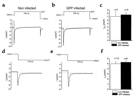
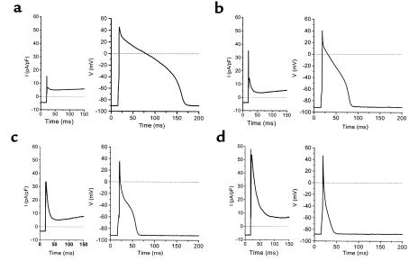
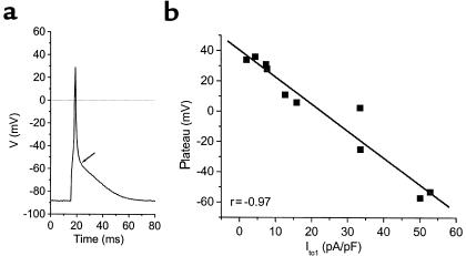
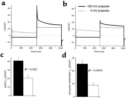
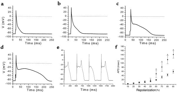
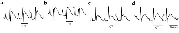
Heart failure leads to marked suppression of the Ca(2+)-independent transient outward current (I(to1)), but it is not clear whether I(to1) downregulation suffices to explain the concomitant action potential prolongation. To investigate the role of I(to1) in cardiac repolarization while circumventing culture-related action potential alterations, we injected adenovirus vectors in vivo to overexpress or to suppress I(to1) in guinea pigs and rats, respectively. Myocytes were isolated 72 hours after intramyocardial injection and stimulation of the ecdysone-inducible vectors with intraperitoneal injection of an ecdysone analog. Kv4.3-infected guinea pig myocytes exhibited robust transient outward currents. Increasing density of I(to1) progressively depressed the plateau potential in Kv4. 3-infected guinea pig myocytes and abbreviated action potential duration (APD). In vivo infection with a dominant-negative Kv4. 3-W362F construct suppressed peak I(to1) in rat ventriculocytes, elevated the plateau height, significantly prolonged the APD, and resulted in a prolongation by about 30% of the QT interval in surface electrocardiogram recordings. These results indicate that I(to1) plays a crucial role in setting the plateau potential and overall APD, supporting a causative role for suppression of this current in the electrophysiological alterations of heart failure. The electrocardiographic findings indicate that somatic gene transfer can be used to create gene-specific animal models of the long QT syndrome.


Figures

Figure 1
Figure 1
L-type calcium currents ICaL and delayed rectifier currents IKr were not different in noninfected guinea pig myocytes compared with myocytes that were in vivo infected with a reporter (GFP) adenovirus. Peak ICaL current density was measured at 0 mV after a prepulse to –40 mV in noninfected (a) and GFP-infected (b) myocytes. IKr tail currents in noninfected cells (d) measured at –100 mV after a 200-ms depolarization step to –10 mV were compared with GFP-infected myocytes (e). Original current traces and mean current densities indicate that adenovirus infection itself did not affect ICaL (ac) and IKr (df).
Figure 2
Figure 2
Confocal fluorescence image of typical in vivo coinfected guinea pig myocytes. Guinea pig myocardium was coinjected with the receptor virus AdCGI-DBEcR and the reporter virus AdE8I-Kv4.3. Seventy-two hours after injection and stimulation with GS-E, myocytes were isolated and CD8 was viewed with R-phycoerythrin–conjugated CD8 antibodies. The cytosolic green fluorescence of the myocyte verifies infection with the receptor virus. Red staining of the surface membrane indicates induced expression of CD8 from the reporter virus, which also carries the ion channel gene. Infected cells were readily distinguishable from the background autofluorescence of noninfected cells (transmitted light image shown in blue plane).
Figure 3
Figure 3
Transient outward currents and action potentials of 4 different guinea pig myocytes (ad) that were infected in vivo with AdCGI-DBEcR and AdE8I-Kv4.3. Seventy-two hours after injection and stimulation with GS-E, myocytes were isolated. Transient outward currents were elicited by depolarization pulses to +40 mV from a holding potential of –100 mV. In vivo adenovirus-mediated introduction of Ito1 substantially changed the action potential waveform of acutely dissected guinea pig myocytes in an Ito1 density–dependent manner, resulting in a depression of the plateau voltage and abbreviation of the overall APD. (d) Introduction of very large Ito1 densities caused a spikelike action potential.
Figure 4
Figure 4
Effect of Ito1 current size on the height of the action potential plateau in guinea pig myocytes infected in vivo with AdCGI-DBEcR and AdE8I-Kv4.3. (a) Introduction of Ito1 (50.1 pA/pF) into a guinea pig myocyte depressed the voltage of the early plateau phase measured at d2V/dt2=0 (arrow) (at the transition from early repolarization to final repolarization; ref. 27). (b) The suppression of the action potential plateau correlated well with the introduced Ito1 density (n = 10).
Figure 5
Figure 5
In vivo expression of AdE8I-Kv4.3-W362F suppressed Ito1 in adult rat myocytes. Transient outward currents elicited by test pulses to +40 mV in noninfected myocytes (a) are compared with currents in AdE8I-Kv4.3-W362F–infected myocytes (b). Mean values of peak Ito1 (c) and of the difference between the fully primed and prepulse-inactivated (0 mV) (d) currents indicate a significant suppression of native rat cardiac Ito1 in Kv4.3-W362F–infected myocytes (n = 7; filled bars) compared with wild-type (n = 10; shaded bars).
Figure 6
Figure 6
Action potentials of rat cardiomyocytes were prolonged by dominant-negative in vivo Ito1 suppression. Infection of rat myocytes with AdE8I-Kv4.3W362F substantially changed the action potential waveform (c and d) compared with noninfected cells (a and b), resulting in an elevation of the plateau phase and prolongation of the overall APD. In 1 rat myocyte, Ito1 reduction led to a notch-and-dome–shaped action potential waveform (d) and to frequent EADs (e). Mean APDs of wild-type (closed squares; n = 10) and Kv4.3W362F-infected myocytes (closed circles; n = 7) plotted as a function of the percentage of repolarization (f) demonstrate that Ito1 suppression did not exhibit a significant effect on the APD at 10–50% repolarization, but significantly prolonged the APD60 to APD90. Values marked by (A) are significant (P < 0.05).
Figure 7
Figure 7
More widespread infection with AdKv4.3W362F leads to a prolongation of the QT interval. ECG recordings were performed after widespread infection with Kv4.3-W362F or GFP (controls). ECGs revealed a 29.8 ± 6.0% prolongation of the QT interval (P = 0.02; n = 3) in Kv4.3-W362F-infected rats 72 hours after intramyocardial injection (d; note the extension of the QT interval into the P wave) compared with immediate postoperative recordings (c), consistent with the marked prolongation of ventricular action potentials. No change in the QT interval was observed in GFP-infected control animals postoperatively (a) compared with 72 hours after infection (b).

References

    1. CONSENSUS Trail Study Group. Effects of enalapril on mortality in severe congestive heart failure: results of the Cooperative North Scandinavian Enalapril Survival Study (CONSENSUS) N Engl J Med. 1987;316:1429–1435. - PubMed
    1. SOLVD Investigators. Effect of enalapril on survival in patients with reduced left ventricular ejection fractions and congestive heart failure. N Engl J Med. 1991;325:293–302. - PubMed
    1. CIBIS InvestigatorsCommittees. A randomized trial of beta-blockade in heart failure. The Cardiac Insufficiency Bisoprolol Study (CIBIS) Circulation. 1994;90:1765–1773. - PubMed
    1. Torp-Pedersen C, et al. Dofetilide in patients with congestive heart failure and left ventricular dysfunction. Danish Investigations of Arrhythmia and Mortality on Dofetilide Study Group. N Engl J Med. 1999;341:857–865. - PubMed
    1. Beuckelmann DJ, Näbauer M, Erdmann E. Alterations of K+ currents in isolated human ventricular myocytes from patients with terminal heart failure. Circ Res. 1993;73:379–385. - PubMed

Publication types