. 2000 May 15;28(10):2049-59.
doi: 10.1093/nar/28.10.2049.

Domain specific interaction in the XRCC1-DNA polymerase beta complex

Affiliations

Domain specific interaction in the XRCC1-DNA polymerase beta complex

A Marintchev et al. Nucleic Acids Res. .

Abstract

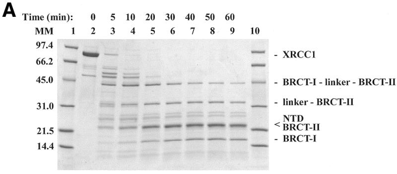
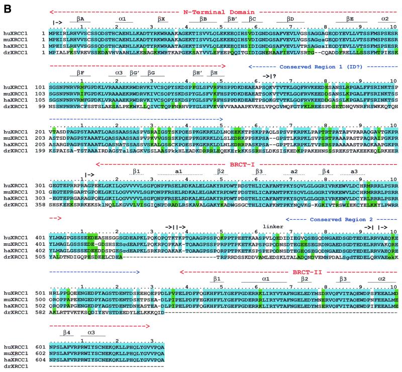
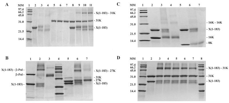
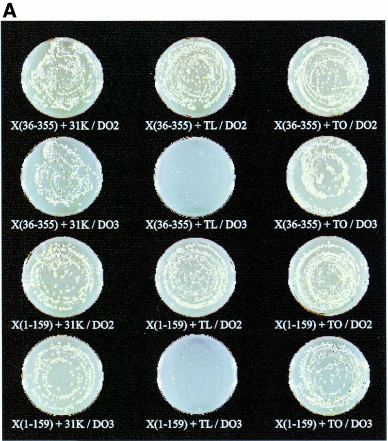
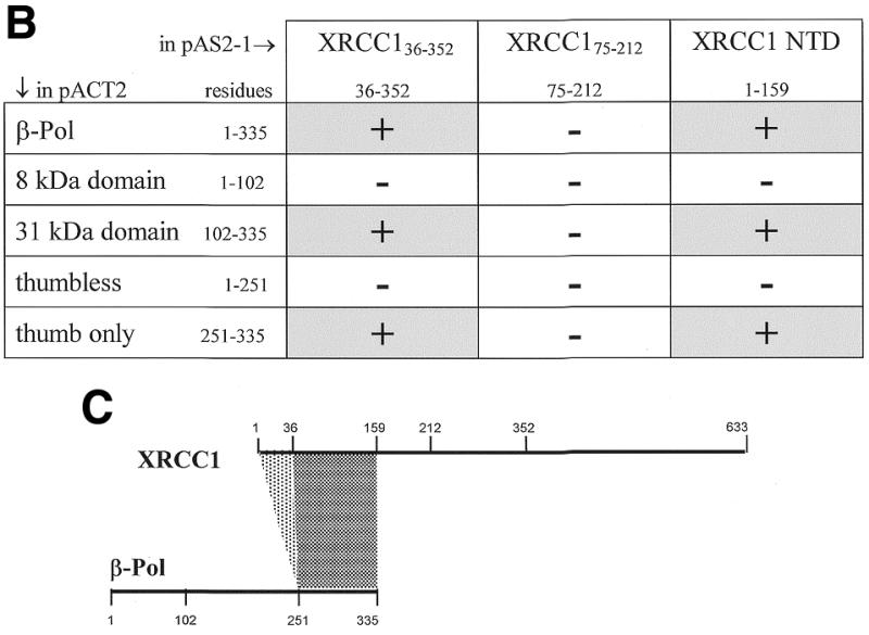
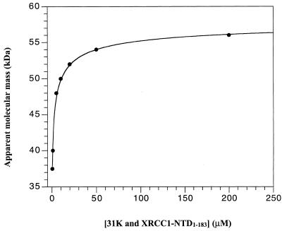
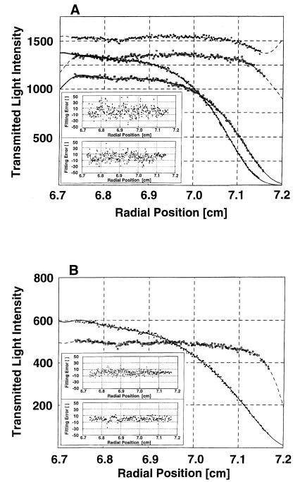
XRCC1 (X-ray cross-complementing group 1) is a DNA repair protein that forms complexes with DNA polymerase beta (beta-Pol), DNA ligase III and poly-ADP-ribose polymerase in the repair of DNA single strand breaks. The domains in XRCC1 have been determined, and characterization of the domain-domain interaction in the XRCC1-beta-Pol complex has provided information on the specificity and mechanism of binding. The domain structure of XRCC1, determined using limited proteolysis, was found to include an N-terminal domain (NTD), a central BRCT-I (breast cancer susceptibility protein-1) domain and a C-terminal BRCT-II domain. The BRCT-I-linker-BRCT-II C-terminal fragment and the linker-BRCT-II C-terminal fragment were relatively stable to proteolysis suggestive of a non-random conformation of the linker. A predicted inner domain was found not to be stable to proteolysis. Using cross-linking experiments, XRCC1 was found to bind intact beta-Pol and the beta-Pol 31 kDa domain. The XRCC1-NTD(1-183)(residues 1-183) was found to bind beta-Pol, the beta-Pol 31 kDa domain and the beta-Pol C-terminal palm-thumb (residues 140-335), and the interaction was further localized to XRCC1-NTD(1-157)(residues 1-157). The XRCC1-NTD(1-183)-beta-Pol 31 kDa domain complex was stable at high salt (1 M NaCl) indicative of a hydrophobic contribution. Using a yeast two-hybrid screen, polypeptides expressed from two XRCC1 constructs, which included residues 36-355 and residues 1-159, were found to interact with beta-Pol, the beta-Pol 31 kDa domain, and the beta-Pol C-terminal thumb-only domain polypeptides expressed from the respective beta-Pol constructs. Neither the XRCC1-NTD(1-159), nor the XRCC1(36-355)polypeptide was found to interact with a beta-Pol thumbless polypeptide. A third XRCC1 polypeptide (residues 75-212) showed no interaction with beta-Pol. In quantitative gel filtration and analytical ultracentrifugation experiments, the XRCC1-NTD(1-183)was found to bind beta-Pol and its 31 kDa domain in a 1:1 complex with high affinity (K(d) of 0.4-2.4 microM). The combined results indicate a thumb-domain specific 1:1 interaction between the XRCC1-NTD(1-159)and beta-Pol that is of an affinity comparable to other binding interactions involving beta-Pol.


Figures

Figure 1
Figure 1
Limited tryptic digestion of XRCC1. (A) SDS–PAGE analysis of the tryptic digestion using 0.2 mg/ml of XRCC1 and 0.2 µg/ml of trypsin. Lanes 1 and 10, molecular mass markers; lane 2, undigested purified XRCC1; and lanes 3–9, digestion times as indicated. The identity of the tryptic fragments is indicated on the right. (B) Alignment of XRCC1 sequences from human (huXRCC1, SwissProt accession no. P18887), mouse (muXRCC1, SwissProt accession no. Q60596), hamster (haXRCC1, GenBank accession no. AAC40038) and Drosophila (drXRCC1, GenBank accession no. AAD33589). Identities in at least three sequences are highlighted in cyan and similarities are highlighted in green. The numbering over the sequences is based on huXRCC1. Insertions with respect to huXRCC1 in the other sequences are not shown and their positions are indicated with pairs of lower case letters. The conserved regions are indicated above the sequence. The known secondary structure elements for NTD and BRCT-II are marked with a solid line and the secondary structure elements for BRCT-I, predicted based on homology to BRCT-II, are marked with a dotted line. The start and expected end of a tryptic fragment (A) are indicated with │-> and ->│, respectively.
Figure 1
Figure 1
Limited tryptic digestion of XRCC1. (A) SDS–PAGE analysis of the tryptic digestion using 0.2 mg/ml of XRCC1 and 0.2 µg/ml of trypsin. Lanes 1 and 10, molecular mass markers; lane 2, undigested purified XRCC1; and lanes 3–9, digestion times as indicated. The identity of the tryptic fragments is indicated on the right. (B) Alignment of XRCC1 sequences from human (huXRCC1, SwissProt accession no. P18887), mouse (muXRCC1, SwissProt accession no. Q60596), hamster (haXRCC1, GenBank accession no. AAC40038) and Drosophila (drXRCC1, GenBank accession no. AAD33589). Identities in at least three sequences are highlighted in cyan and similarities are highlighted in green. The numbering over the sequences is based on huXRCC1. Insertions with respect to huXRCC1 in the other sequences are not shown and their positions are indicated with pairs of lower case letters. The conserved regions are indicated above the sequence. The known secondary structure elements for NTD and BRCT-II are marked with a solid line and the secondary structure elements for BRCT-I, predicted based on homology to BRCT-II, are marked with a dotted line. The start and expected end of a tryptic fragment (A) are indicated with │-> and ->│, respectively.
Figure 2
Figure 2
Glutaraldehyde (GA) cross-linking of XRCC1-NTD1–183 designated as X(1–183) with full-length β-Pol and β-Pol domains. β-Pol domains included the N-terminal 8 kDa domain and the C-terminal 31 kDa domain. The 27 kDa palm–thumb domain is a C-terminal proteolysis product derived from the 31 kDa domain. The 16 kDa domain includes the 8 kDa domain and the fingers domain of the 31 kDa domain. Cross-linking was performed for 30 min, unless otherwise stated. The concentrations of the proteins were as follows: X(1–183), 20 µM; β-Pol, 4 µM; β-Pol 31 kDa domain (31K), 10 µM; β-Pol 27 kDa fragment (27K), <10 µM (Materials and Methods); β-Pol 16 kDa fragment (16K), 40 µM; β-Pol 8 kDa domain (8K), 40 µM. The reactions were stopped by boiling in SDS sample buffer and the proteins were separated on 15% acrylamide gels. Gels were stained with Coomassie blue. (A) Lane 1, molecular mass markers; lane 2, X(1–183); lane 3, X(1–183) + GA, 10 min; lane 4, X(1–183) + GA, 30 min; lane 5, 31K; lane 6, 31K + GA, 10 min; lane 7, 31K + GA, 30 min; lane 8, X(1–183) + 31K; lane 9, X(1–183) + 31K + GA, 10 min; lane 10, X(1–183) + 31K + GA, 20 min; lane 11, X(1–183) + 31K + GA, 30 min. (Note: the band of free X(1–183) doubles upon modification by GA.) (B) Lane 1, X(1–183) + β-Pol; lane 2, X(1–183) + β-Pol + GA; lane 3, β-Pol + GA; lane 4, molecular mass markers; lane 5, X(1–183) + 27K; lane 6, X(1–183) + 27K + GA; lane 7, 27K + GA. (C) Lane 1, molecular mass markers; lane 2, X(1–183) +16K; lane 3, X(1–183) + 16 K + GA; lane 4, 16K + GA; lane 5, X(1–183) + 8K; lane 6, X(1–183) + 8K + GA; lane 7, 8K + GA. (D) Effect of salt on cross-linking of XRCC1-NTD1–183 [X(1–183)] with β-Pol 31 kDa domain (31K). Lanes 1 and 8, molecular mass markers; lane 2, X(1–183) + 31K; lanes 3–7, X(1–183) + 31K + GA in the following salt concentrations: 0 mM NaCl (lane 3); 100 mM NaCl (lane 4); 500 mM NaCl (lane 5); 1 M NaCl (lane 6); 2 M NaCl (lane 7).
Figure 3
Figure 3
Yeast two-hybrid interactions between regions of XRCC1 and β-Pol. (A) Yeast CG1945 cells co-transformed with the indicated XRCC1 BD and β-Pol AD constructs and grown on DO2 and DO3 plates. Yeast colonies of at least 1 mm were visible 3 days after co-transformation. The designations are as follows: XRCC136–355 [X(36–355)]; XRCC1-NTD1–159 [X(1–159)]; β-Pol 31 kDa domain, Arg102 to the C-terminal Glu335 (31K); β-Pol thumbless, Met1 to Asp251 (TL); β-Pol thumb-only, Asp251 to the C-terminal Glu335 (TO). DO2 is defined media lacking Trp and Leu. Visible colonies on DO2 plates indicate transformation with the BD and AD two-hybrid vector plasmids. DO3 is defined media lacking Trp, Leu and His with HIS3 inhibitor 3AT added to a final concentration of 25 mM. Visible colonies on DO3 plates report protein interactions. (B) Summary of the interactions observed between the XRCC1 BD and β-Pol AD expressed proteins. (C) Summary of the polypeptide segments that displayed a yeast two-hybrid interaction.
Figure 3
Figure 3
Yeast two-hybrid interactions between regions of XRCC1 and β-Pol. (A) Yeast CG1945 cells co-transformed with the indicated XRCC1 BD and β-Pol AD constructs and grown on DO2 and DO3 plates. Yeast colonies of at least 1 mm were visible 3 days after co-transformation. The designations are as follows: XRCC136–355 [X(36–355)]; XRCC1-NTD1–159 [X(1–159)]; β-Pol 31 kDa domain, Arg102 to the C-terminal Glu335 (31K); β-Pol thumbless, Met1 to Asp251 (TL); β-Pol thumb-only, Asp251 to the C-terminal Glu335 (TO). DO2 is defined media lacking Trp and Leu. Visible colonies on DO2 plates indicate transformation with the BD and AD two-hybrid vector plasmids. DO3 is defined media lacking Trp, Leu and His with HIS3 inhibitor 3AT added to a final concentration of 25 mM. Visible colonies on DO3 plates report protein interactions. (B) Summary of the interactions observed between the XRCC1 BD and β-Pol AD expressed proteins. (C) Summary of the polypeptide segments that displayed a yeast two-hybrid interaction.
Figure 4
Figure 4
Determination of the Kd (0.43 µM) for the binding of XRCC1-NTD1–183 to the 31 kDa domain of β-Pol (31K) by gel filtration HPLC. The proteins were mixed at 1:1 molar ratio with concentrations ranging from 0.5 to 200 µM. XRCC1-NTD1–183 and 31K migrated as a single chromatographic peak throughout the entire range of concentrations and the complex displayed a concentration-dependent average molecular mass. The apparent molecular mass of the complex, XRCC11–183–31K, calculated from the retention time was plotted as a function of the concentration. The Kd of 0.43 ± 0.05 µM was fitted as described in Materials and Methods.
Figure 5
Figure 5
Analytical ultracentrifugation analysis of the interaction of XRCC1-NTD1–183 with full-length β-Pol and its 31 kDa domain. (A) Global curve-fitting of transmitted light intensity data at 4°C from two centrifuge cells loaded with different total concentrations of XRCC1-NTD1–183 and full-length β-Pol, in a 1:1 molar ratio, using the mathematical model given in equation 6 (Materials and Methods). The top two curves represent the reference channels and the bottom two the corresponding sample channels. The data points (open squares) correspond to the cells loaded with concentrations of ~9 µM of each protein while data points shown by stars correspond to the cells loaded with 16 µM of each protein. For each sample concentration, the sample and reference data curves and the fitting error plots have the same symbol. The quality of fit is excellent as can be seen in the inset, which shows the fitting errors for the two sample channels. (B) Curve fitting of transmitted light intensity data from a single centrifuge cell containing XRCC1-NTD1–183 and the β-Pol 31 kDa domain, in a 1:1 ratio, at 20°C, using the mathematical model given in equation 6. The concentration for each protein was ~15 µM. The data points for the reference channel are shown as (open squares), and the data points for the sample channel are shown by triangles. The quality of fit is excellent as can be seen in the inset, which shows the fitting errors. The top error plot represents the sample and the bottom the reference channel. The latter was fitted using cubic splines.

Similar articles

Cited by

References

    1. Thompson L.H., Brookman,K.W., Jones,N.J., Allen,S.A. and Carrano,A.V. (1990) Mol. Cell. Biol., 10, 6160–6171. - PMC - PubMed
    1. Tebbs R.S., Flannery,M.L., Meneses,J.J., Hartmann,A., Tucker,J.D., Thompson,L.H., Cleaver,J.E. and Pedersen,R.A. (1999) Dev. Biol., 208, 513–529. - PubMed
    1. Sobol R.W., Horton,J.K., Kuhn,R., Gu,H., Singhal,R.K., Prasad,R., Rajewsky,K. and Wilson,S.H. (1996) Nature, 379, 183–186. - PubMed
    1. Thompson L.H. and West,M.G. (2000) Mutat. Res., 459, 1–18. - PubMed
    1. Kubota Y., Nash,R.A., Klungland,A., Schar,P., Barnes,D.E. and Lindahl,T. (1996) EMBO J., 15, 6662–6670. - PMC - PubMed

Publication types

MeSH terms