. 2000 May 15;28(10):2114-21.
doi: 10.1093/nar/28.10.2114.

Structural organization of Staf-DNA complexes

Affiliations

Structural organization of Staf-DNA complexes

M Schaub et al. Nucleic Acids Res. .

Abstract

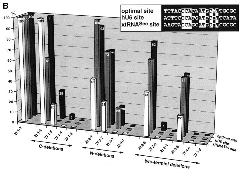
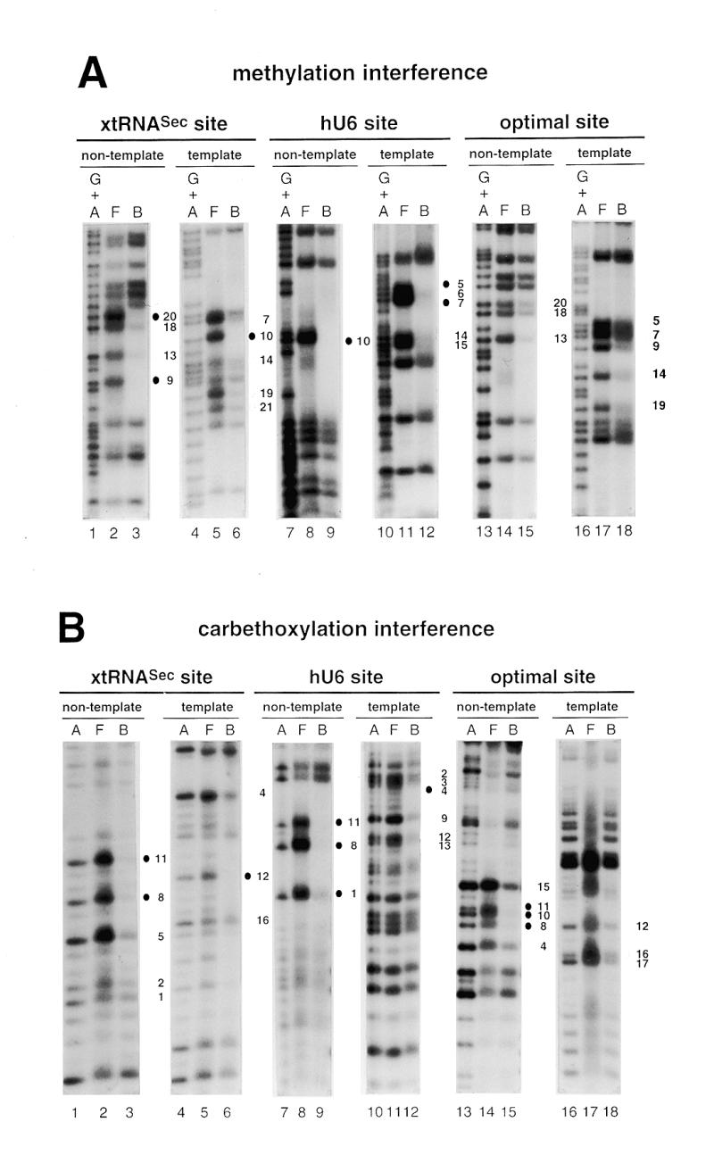
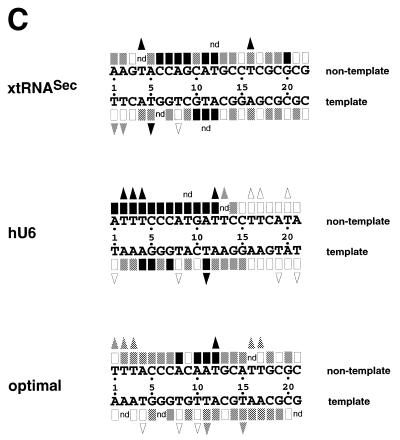
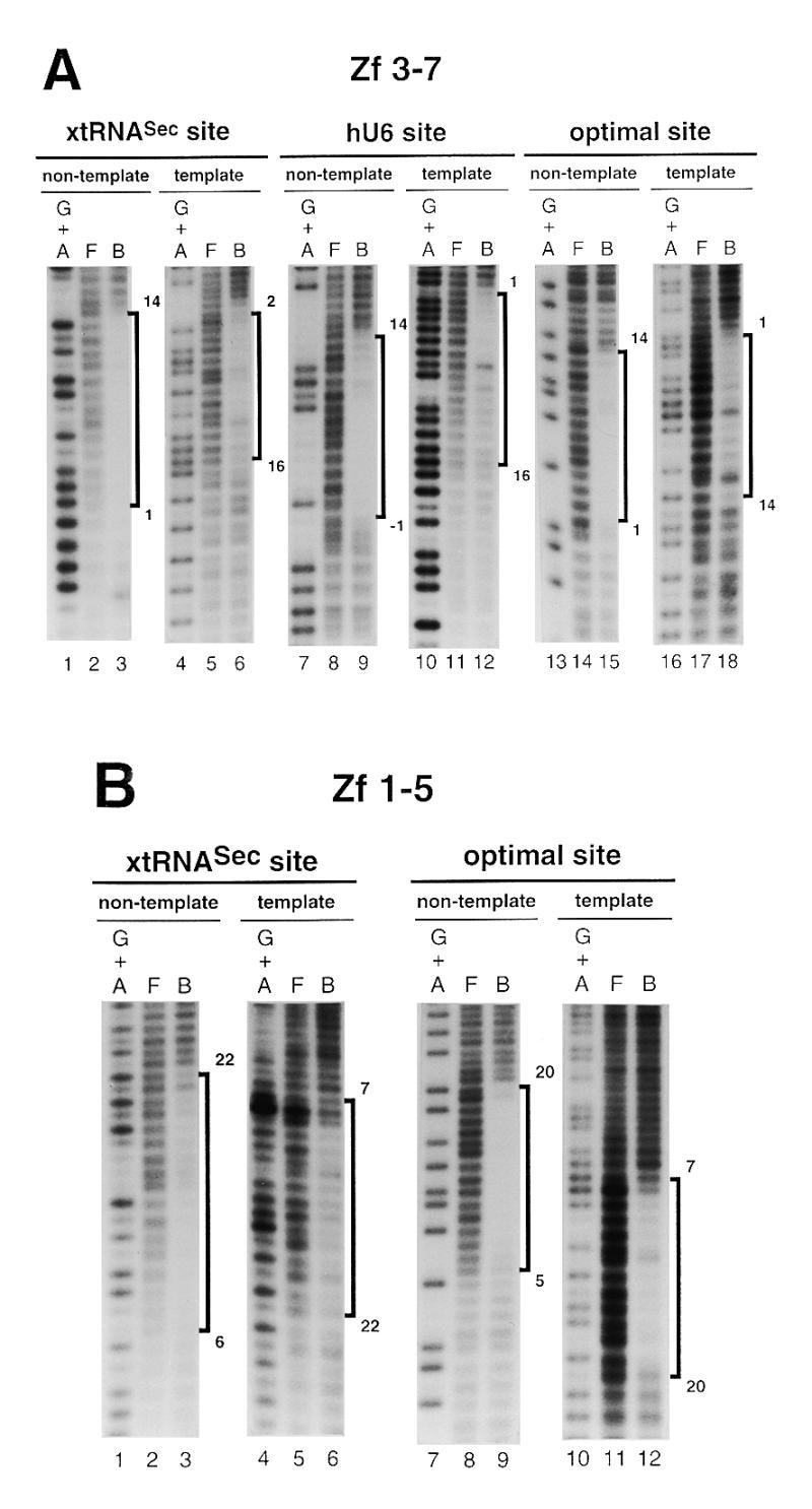
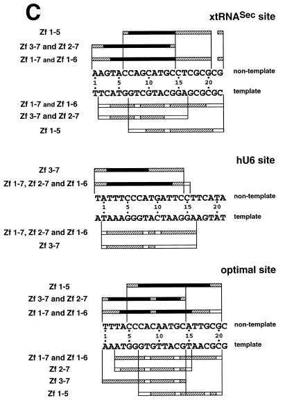
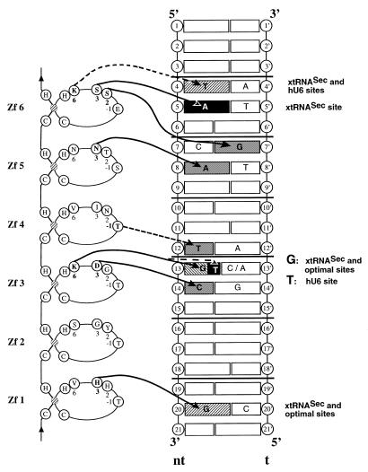
The transactivator Staf, which contains seven contiguous zinc fingers of the C(2)-H(2)type, exerts its effects on gene expression by binding to specific targets in vertebrate small nuclear RNA (snRNA) and snRNA-type gene promoters. Here, we have investigated the interaction of the Staf zinc finger domain with the optimal Xenopus selenocysteine tRNA (xtRNA(Sec)) and human U6 snRNA (hU6) Staf motifs. Generation of a series of polypeptides containing increasing numbers of Staf zinc fingers tested in binding assays, by interference techniques and by binding site selection served to elucidate the mode of interaction between the zinc fingers and the Staf motifs. Our results provide strong evidence that zinc fingers 3-6 represent the minimal zinc finger region for high affinity binding to Staf motifs. Furthermore, we show that the binding of Staf is achieved through a broad spectrum of close contacts between zinc fingers 1-6 and xtRNA(Sec)or optimal sites or between zinc fingers 3-6 and the hU6 site. Extensive DNA major groove contacts contribute to the interaction with Staf that associates more closely with the non-template than with the template strand. Based on these findings and the structural information provided by the solved structures of other zinc finger-DNA complexes, we propose a model for the interaction between Staf zinc fingers and the xtRNA(Sec), optimal and hU6 sites.


Figures

Figure 1
Figure 1
Binding of wild-type and truncated Staf zinc finger domains to the optimal Staf-binding site and to the Staf motifs in the human snRNA U6 (hU6 site) and in the X.laevis tRNASec (xtRNASec site) promoters. (A) Amino acid sequence of residues 255–476 displaying the sequence alignments of the seven zinc fingers (18). Gaps (<) have been introduced at two locations to maximize the match. Cysteines, histidines and invariant hydrophobic residues are depicted in bold. Open and solid triangles indicate the N- and the C-termini of the various Staf zinc finger domains expressed as GST fusion proteins in E.coli, respectively. (B) Relative binding efficiencies of wild-type and truncated Staf zinc finger domains for the optimal, hU6 and xtRNASec sites. The histogram plots the amount of probe bound to the truncated proteins, relative to that obtained with the wild-type zinc finger domain Zf 1–7. Binding reactions contained 2 nM DNA and 60 nM fusion protein. The results of one representative experiment for each protein and probe are shown. Two other independent determinations gave similar results. Sequence comparisons between the optimal, hU6 and xtRNASec Staf-binding sites used in this study are shown in the upper right pannel. Nucleotide identities between the different elements are boxed.
Figure 1
Figure 1
Binding of wild-type and truncated Staf zinc finger domains to the optimal Staf-binding site and to the Staf motifs in the human snRNA U6 (hU6 site) and in the X.laevis tRNASec (xtRNASec site) promoters. (A) Amino acid sequence of residues 255–476 displaying the sequence alignments of the seven zinc fingers (18). Gaps (<) have been introduced at two locations to maximize the match. Cysteines, histidines and invariant hydrophobic residues are depicted in bold. Open and solid triangles indicate the N- and the C-termini of the various Staf zinc finger domains expressed as GST fusion proteins in E.coli, respectively. (B) Relative binding efficiencies of wild-type and truncated Staf zinc finger domains for the optimal, hU6 and xtRNASec sites. The histogram plots the amount of probe bound to the truncated proteins, relative to that obtained with the wild-type zinc finger domain Zf 1–7. Binding reactions contained 2 nM DNA and 60 nM fusion protein. The results of one representative experiment for each protein and probe are shown. Two other independent determinations gave similar results. Sequence comparisons between the optimal, hU6 and xtRNASec Staf-binding sites used in this study are shown in the upper right pannel. Nucleotide identities between the different elements are boxed.
Figure 2
Figure 2
Interference with Staf binding by methylation, carbethoxylation, KMnO4 modification and depyrimidation of DNA fragments containing the xtRNASec, hU6 or optimal Staf-binding sites. The DNA fragments were 32P-labeled at their 5′-ends on the template or non-template strands, partially methylated (A) or carbethoxylated (B) and subjected to interference analysis. DMS modification and treatment of the modified DNA were performed under conditions allowing cleavage at only the modified guanine bases. Lanes A and G + A, sequencing reactions; lanes F and B, free and bound DNAs. Bases whose modifications interfered with Staf binding are shown by dark (total interference) or gray (partial interference) circles. Bases are numbered according to the Staf consensus binding site (21). (C) Summary of the interference experiments. The full and partial interference effects of guanine methylation, adenine carbethoxylation and pyrimidine removal on Staf binding are represented by dark and gray squares, respectively. The full and partial interference effects on the binding of Staf by KMnO4 modification of thymines are depicted by dark and gray triangles, respectively. Open squares and triangles show positions where base modifications or removal did not interfere with Staf binding; nd, not determined.
Figure 2
Figure 2
Interference with Staf binding by methylation, carbethoxylation, KMnO4 modification and depyrimidation of DNA fragments containing the xtRNASec, hU6 or optimal Staf-binding sites. The DNA fragments were 32P-labeled at their 5′-ends on the template or non-template strands, partially methylated (A) or carbethoxylated (B) and subjected to interference analysis. DMS modification and treatment of the modified DNA were performed under conditions allowing cleavage at only the modified guanine bases. Lanes A and G + A, sequencing reactions; lanes F and B, free and bound DNAs. Bases whose modifications interfered with Staf binding are shown by dark (total interference) or gray (partial interference) circles. Bases are numbered according to the Staf consensus binding site (21). (C) Summary of the interference experiments. The full and partial interference effects of guanine methylation, adenine carbethoxylation and pyrimidine removal on Staf binding are represented by dark and gray squares, respectively. The full and partial interference effects on the binding of Staf by KMnO4 modification of thymines are depicted by dark and gray triangles, respectively. Open squares and triangles show positions where base modifications or removal did not interfere with Staf binding; nd, not determined.
Figure 3
Figure 3
Hydroxyl radical interference patterns obtained with Staf Zf 1–5 and Zf 3–7 on the xtRNASec, hU6 and optimal sites. The 5′-end-labeled non-template or template strands containing the xtRNASec, hU6 and optimal sites were subjected to hydroxyl radical cleavage. Gapped DNAs were incubated separately with GST-fused Zf 1–5 and Zf 3–7. (A) Missing nucleoside interference patterns obtained on the xtRNASec, hU6 and optimal sites with Zf 3–7 and (B) with Zf 1–5 on the xtRNASec and optimal sites. Lanes G + A, F and B as in Figure 2. (C) Schematic representation of the results with comparison to previous results obtained with Zf 1–7, Zf 1–6 and Zf 2–7 (21,22). Regions of interference are boxed; dark boxes, strongest interference; hatched boxes, moderate interference; open boxes, weakest interference. The base pairs in the Staf-binding site are numbered –1 to 22, starting at the 5′-end of the non-template strand, with reference to the consensus binding site derived by in vitro selection (21).
Figure 3
Figure 3
Hydroxyl radical interference patterns obtained with Staf Zf 1–5 and Zf 3–7 on the xtRNASec, hU6 and optimal sites. The 5′-end-labeled non-template or template strands containing the xtRNASec, hU6 and optimal sites were subjected to hydroxyl radical cleavage. Gapped DNAs were incubated separately with GST-fused Zf 1–5 and Zf 3–7. (A) Missing nucleoside interference patterns obtained on the xtRNASec, hU6 and optimal sites with Zf 3–7 and (B) with Zf 1–5 on the xtRNASec and optimal sites. Lanes G + A, F and B as in Figure 2. (C) Schematic representation of the results with comparison to previous results obtained with Zf 1–7, Zf 1–6 and Zf 2–7 (21,22). Regions of interference are boxed; dark boxes, strongest interference; hatched boxes, moderate interference; open boxes, weakest interference. The base pairs in the Staf-binding site are numbered –1 to 22, starting at the 5′-end of the non-template strand, with reference to the consensus binding site derived by in vitro selection (21).
Figure 4
Figure 4
Models for the binding of Staf to the xtRNASec, optimal and hU6 Staf-binding sites. C2-H2 Zf 1–Zf 6 are represented with the two invariant cysteines, histidines and the bound zinc (hatched). The amino acids at positions –1, 2, 3 and 6 in the zinc finger α-helix that are crucial for making base contacts in the solved structures of zinc finger–DNA complexes are indicated. The DNA helix represents the Staf-binding sites, with bases on the non-template (nt) strand numbered 1–21 according to the consensus Staf-binding site (21) and 1′–21′ on the template strand. Horizontal lines define triplet subsites. Solid and dotted arrows indicate the predicted base contacts based on the solved structures and tentative recognition codes, respectively. Only base pairs involved in the putative zinc finger–DNA contacts are indicated; gray boxes, contacts with the three sites; hatched boxes, contacts with two sites; dark boxes, contact with one site. The relevant sites are indicated on the right.

Similar articles

Cited by

References

    1. Miller J., McLachlan,A.D. and Klug,A. (1985) EMBO J., 4, 1609–1614. - PMC - PubMed
    1. Rosenberg U.B., Schröder,C., Preiss,A., Kienlin,A., Cote,S., Riede,I. and Jäckle,H. (1986) Nature, 319, 336–339.
    1. Tautz D., Lehmann,R., Schnürch,H., Schuh,R., Seifert,E., Kienlin,A., Jones,K. and Jäckle,H. (1987) Nature, 327, 383–389.
    1. Turner J. and Crossley,M. (1999) Trends Biochem. Sci., 24, 236–240. - PubMed
    1. Schuh R., Aicher,W., Gaul,U., Côté,S., Preiss,A., Maier,D., Seifert,E., Nauber,U., Schröder,C., Kemler,R. and Jäckle,H. (1986) Cell, 47, 1025–1032. - PubMed

Publication types

MeSH terms