

Review
doi: 10.1093/emboj/19.8.1745.
Transcriptional control by the TGF-beta/Smad signaling system
Affiliations
- PMID: 10775259
- PMCID: PMC302010
- DOI: 10.1093/emboj/19.8.1745
Item in Clipboard
Review
Transcriptional control by the TGF-beta/Smad signaling system
EMBO J.
.
No abstract available
Figures
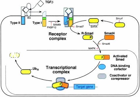
Fig. 1. Schematic representation of the TGF-β/Smad signaling engine. This system involves a family of membrane receptor protein kinases and a family of receptor substrates (the Smad proteins) that march into the nucleus where they act as transcription factors. The ligand TGF-β assembles a receptor complex that activates Smads, and the Smads assemble multisubunit complexes that regulate transcription. Two general steps suffice to carry the hormonal stimulus to target genes. The central components of this signaling system are indicated along with the sites of action of various positive and negative regulators. See the text for further details.
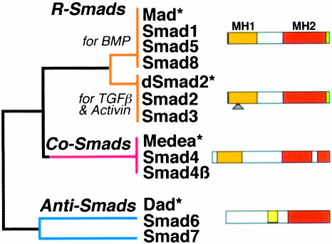
Fig. 2. The Smad family. Simplified dendrogram of sequence similarity between the three Smad subfamilies. The receptor-regulated Smads (R-Smads) and their cooperating Smads (Co-Smads) contain conserved N-terminal (MH1) and C-terminal (MH2) domains separated by a divergent region. Only the MH2 domain is conserved in the inhibitory Smads (Anti-Smads). The green sliver represents the receptor phosphorylation sites at the extreme C-terminus of the R-Smads. The triangle represents the alternatively spliced insert in Smad2. Asterisks denote representative members from Drosophila.
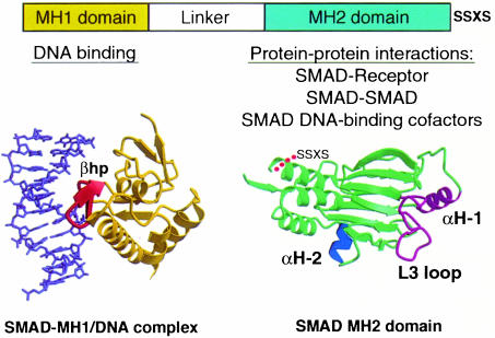
Fig. 3. Smad structural domains and their functions. Representation of the three-dimensional structures of the Smad3 MH1 domain bound to the AGAC sequence, and the Smad2 MH2 domain. The principal interactions of these two domains are listed. The structures involved in these interactions are shown in different colors: the β-hairpin (βhp) that mediated DNA binding, the L3 loop and α-helix 1 (αH-1) that specify Smad interactions with type I receptors, and the α-helix 2 (αH-2) that specifies Smad2 interaction with FAST. SSXS, receptor phosphorylation sites (adapted from Shi et al., 1997, 1998; Wu et al., 2000).
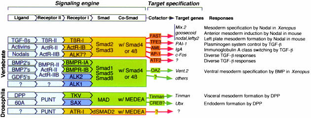
Fig. 4. Making choices through the Smad system.The combinatorial organization of this system as presently understood.
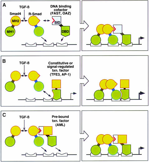
Fig. 5. Smad transcriptional partners. General models for the recognition and regulation of specific target genes by Smads in concert with DNA-binding adaptors such as FAST and OAZ (model A) and constitutive (e.g. TFE and CBF) or signal-regulated (e.g. AP-1) transcription factors that interact with the MH1 domain upon agonist activation (model B) or with the MH2 domain in the basal state (model C). MH1 and MH2, Smad domains; orange boxes, transactivator domains; SID, Smad interaction domain. Although two Smad DNA sites are depicted in each model, only one may be used in certain response elements.
References
-
- Akiyoshi S., Inoue,H., Hanai,J., Kusanagi,K., Nemoto,N., Miyazono,K. and Kawabata,M. (1999) c-Ski acts as a transcriptional co-repressor in transforming growth factor-β signaling through interaction with smads. J. Biol. Chem., 274, 35269–35277. - PubMed
-
- Ashcroft G.S. et al. (1999) Mice lacking Smad3 show accelerated wound healing and an impaired local inflammatory response. Nature Cell Biol., 1, 260–266. - PubMed
-
- Beckmann H., Su,L.K. and Kadesch,T. (1990) TFE3: a helix–loop–helix protein that activates transcription through the immunoglobulin enhancer muE3 motif. Genes Dev., 4, 167–179. - PubMed
-
- Bertolino E., Reimund,B., Wildt-Perinic,D. and Clerc,R. (1995) A novel homeobox protein which recognizes a TGT core and functionally interferes with a retinoid-responsive motif. J. Biol. Chem., 270, 31178–31188. - PubMed
-
- Brown C.B., Boyer,A.S., Runyan,R.B. and Barnett,J.V. (1999) Requirement of type III TGF-β receptor for endocardial cell transformation in the heart. Science, 283, 2080–2082. - PubMed
Publication types
MeSH terms
Substances
LinkOut - more resources
Full Text Sources
Other Literature Sources
Molecular Biology Databases
