Comparative Study
. 2000 Apr 17;19(8):1766-76.
doi: 10.1093/emboj/19.8.1766.

Structure and mechanism of the aberrant ba(3)-cytochrome c oxidase from thermus thermophilus

Affiliations
Comparative Study

Structure and mechanism of the aberrant ba(3)-cytochrome c oxidase from thermus thermophilus

T Soulimane et al. EMBO J. .

Abstract

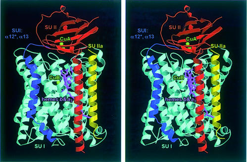
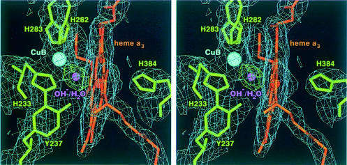
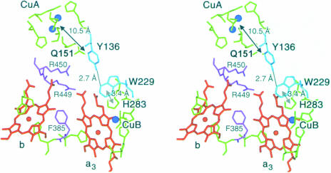
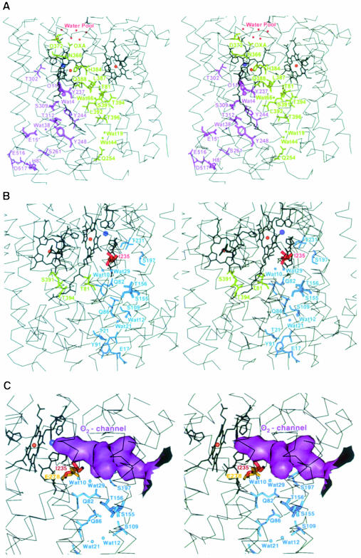
Cytochrome c oxidase is a respiratory enzyme catalysing the energy-conserving reduction of molecular oxygen to water. The crystal structure of the ba(3)-cytochrome c oxidase from Thermus thermophilus has been determined to 2.4 A resolution using multiple anomalous dispersion (MAD) phasing and led to the discovery of a novel subunit IIa. A structure-based sequence alignment of this phylogenetically very distant oxidase with the other structurally known cytochrome oxidases leads to the identification of sequence motifs and residues that seem to be indispensable for the function of the haem copper oxidases, e.g. a new electron transfer pathway leading directly from Cu(A) to Cu(B). Specific features of the ba(3)-oxidase include an extended oxygen input channel, which leads directly to the active site, the presence of only one oxygen atom (O(2-), OH(-) or H(2)O) as bridging ligand at the active site and the mainly hydrophobic character of the interactions that stabilize the electron transfer complex between this oxidase and its substrate cytochrome c. New aspects of the proton pumping mechanism could be identified.


Figures

None
Fig. 1. Stereo ribbon plot of the ba3-cytochrome c oxidase from T.thermophilus viewed parallel to the membrane. The 12 transmembrane helices of subunit I that are also present in the other oxidases are shown in cyan, and the additional transmembrane segment (helices α12* and α13) in dark blue. Subunit II is represented in red, and the new subunit IIa, which corresponds to the first transmembrane helix of subunit II of other oxidases, in yellow. The haem prosthetic groups are depicted as stick models in purple, and the copper atoms as green balls. This figure was created with MOLSCRIPT (Kraulis, 1991) and rendered with RASTER3D (Merritt and Murphy, 1994).
None
Fig. 2. Stereo representation of the binuclear centre haem as3 CuB including the final 2FobsFcalc electron density map contoured at 1.0σ (blue). Haem as3, the histidine ligands and the covalently linked Tyr237 are shown as stick models in orange and green, respectively. The covalent bond between Tyr237 and His233 is well defined in the electron density. The FobsFcalc difference electron density (contoured at 5.0σ, green) between the haem as3 iron and CuB (cyan) is of almost spherical shape and is best interpreted as one oxygen atom (O2–, OH or H2O; purple), located equidistant between the two metal atoms. This figure was prepared with MAIN (Turk, 1992).
None
Fig. 3. Structure-based sequence alignment of subunits I (A) and II (B) of the T.thermophilus ba3-oxidase with the aa3-oxidases from P.denitrificans and bovine heart as well as the engineered soluble CuA-binding domain of the quinol oxidase from E.coli. The transmembrane helices are numbered α1–13 for subunit I and α1 for subunit II. Short connecting helices of subunit I are marked with an asterisk (*) and the β-sheets of subunit II are numbered β1–10. The alignment is shown only for those residues that occupy comparable positions in space (∼60%), and the quality of this structural alignment is indicated with dark and light blue bars for residues at almost identical positions (r.m.s.d. of these Cα atoms 1.1 Å) and those that can be identified as being at similar positions within the structure (r.m.s.d. of these Cα atoms 2.0 Å), respectively. All other residues show no structural similarity between the T.thermophilus ba3-oxidase and other cytochrome oxidases. Strictly conserved amino acids are shown in red, and those with similar chemical characteristics in yellow. This figure was created with ALSCRIPT (Barton, 1993).
None
Fig. 4. Solid surface representation of the electrostatic potential of the proposed cytochrome c-binding surface near the mixed valence CuA site of cytochrome c oxidases (left) and the corresponding front face of cytochrome c (right) for (A) the T.thermophilus ba3-oxidase and its substrate cytochrome c552 (Than et al., 1997) and (B) the bovine heart aa3-oxidase (Tsukihara et al., 1996) and horse heart cytochrome c (Bushnell et al., 1990). Colouring is according to the calculated electrostatic potential of the oxidized oxidases and the reduced cytochromes, and contoured from –40 kT/e (intense red) to 40 kT/e (intense blue). This figure was prepared with GRASP (Nicholls et al., 1993).
None
Fig. 5. Stereo representation of the electron transfer pathways in the ba3-cytochrome c oxidase from T.thermophilus. The haems b and as3 are shown in orange, the copper atoms in blue and the metal-ligating amino acid residues in green. The two arginine residues (Arg450 and Arg449) and Phe385, which are involved in the electron transfer from CuA via haem b to the active site haem as3 CuB are depicted in purple. The residues that form the newly postulated electron transfer pathway leading from CuA to the aromatic ring system of Tyr136 and from there via a hydrogen bond and Trp229 to the CuB ligand His238 are represented in cyan, including the corresponding distances. This figure was prepared with MAIN (Turk, 1992).
None
Fig. 6. Stereo representation of the proton pathways, the O2 channel and the water pool in the ba3-cytochrome c oxidase from T.thermophilus including the Cα traces of the transmembrane helices (thin black lines), the haems and metal ligands (thick black lines) as well as the metal atoms as red and blue balls for the haem iron and copper atoms, respectively. Residues that belong to subunit II are marked by an asterisk. (A) The new ‘Q-pathway’ leading from the cytoplasmic side of the membrane via the haem as3 coordinating His384 to the water pool on top of the haem propionates is represented in green, and the pathway that corresponds to the classical ‘K-pathway’ is shown in purple. (B) Residues of the proton pathway, which corresponds to the classical ‘D-pathway’, are shown in blue, Thr81 and Thr394 that may connect it to Ser391 of the ‘Q-pathway’ are in green and Ile235 in red. (C) Solid surface representation of the O2 input channel (purple), which extends towards the active site including the upper part of the ‘D-pathway’(blue) and Ile235 (red). The highly conserved amino acid Glu278P (yellow), which is indispensable for the proton pumping activity of other oxidases, occupies there the same space as Ile235. This figure was prepared with MAIN (Turk, 1992).

References

    1. Adams M.W.W. and Kelly,R.M. (1998) Finding and using hyperthermophilic enzymes. Trends Biotechnol., 16, 329–332. - PubMed
    1. Barton G.J. (1993) ALSCRIPT: a tool to format multiple sequence alignments. Protein Eng., 6, 37–40. - PubMed
    1. Brünger A.T. et al. (1998) Crystallography and NMR system: a new software suite for macromolecular structure determination. Acta Crystallogr. D, 54, 905–921. - PubMed
    1. Buse G., Soulimane,T., Dewor,M., Meyer,H.E. and Blüggel,M. (1999) Evidence for a copper coordinated histidine–tyrosine crosslink in the active site of cytochrome oxidase. Protein Sci., 8, 985–990. - PMC - PubMed
    1. Bushnell G.W., Louie,G.V. and Brayer,G.D. (1990) High-resolution three-dimensional structure of horse heart cytochrome c. J. Mol. Biol., 214, 585–595. - PubMed

Publication types

MeSH terms