. 2000 Apr 17;19(8):1803-15.
doi: 10.1093/emboj/19.8.1803.

The role of the sid1p kinase and cdc14p in regulating the onset of cytokinesis in fission yeast

Affiliations

The role of the sid1p kinase and cdc14p in regulating the onset of cytokinesis in fission yeast

D A Guertin et al. EMBO J. .

Abstract

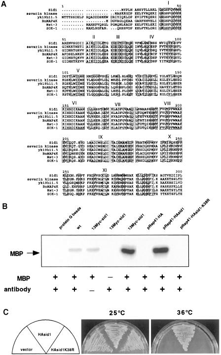
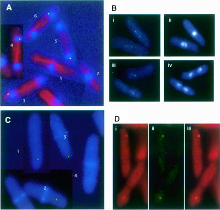
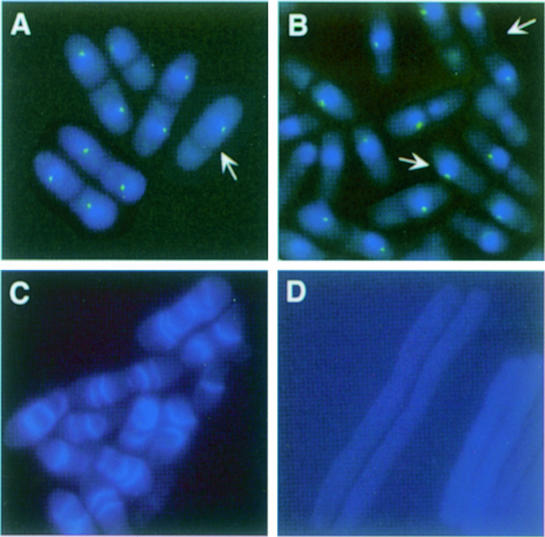
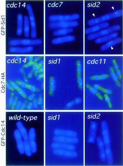
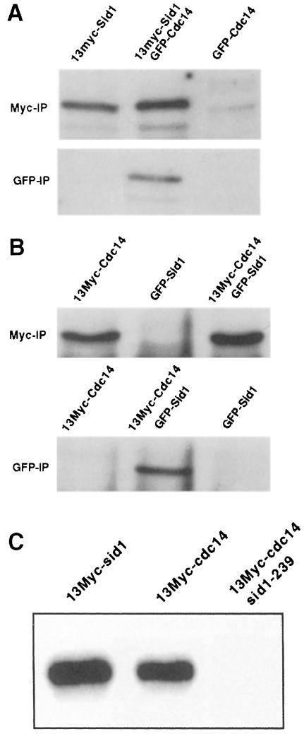
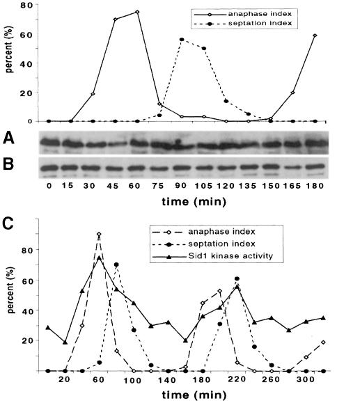
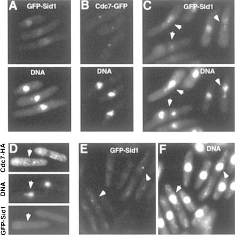
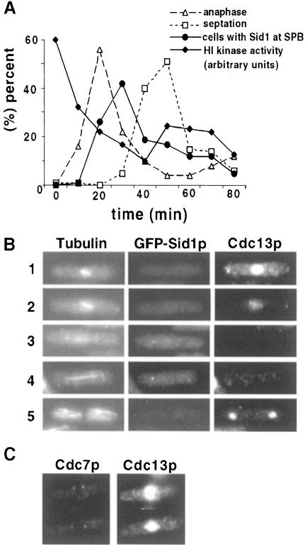
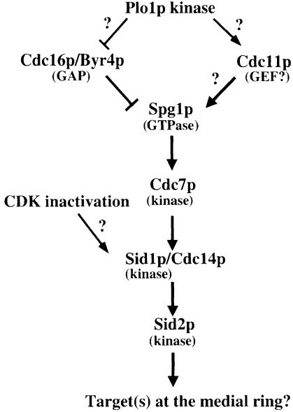
Coordination of mitosis and cytokinesis is crucial for ensuring proper chromosome segregation and genomic stability. In Schizosaccharomyces pombe, the sid genes (cdc7, cdc11, cdc14, spg1, sid1, sid2 and sid4) define a signaling pathway that regulates septation and cytokinesis. Here we describe the characterization of a novel protein kinase, Sid1p. Sid1p localizes asymmetrically to one spindle pole body (SPB) in anaphase. Sid1p localization is maintained during medial ring constriction and septum synthesis and disappears prior to cell separation. Additionally, we found that Cdc14p is in a complex with Sid1p. Epistasis analysis places Sid1p-Cdc14p downstream of Spg1p-Cdc7p but upstream of Sid2p. Finally, we show that cyclin proteolysis during mitosis is unaffected by inactivating the sid pathway; in fact, loss of Cdc2-cyclin activity promotes Sid1p-Cdc14p association with the SPB, possibly providing a mechanism that couples cytokinesis with mitotic exit.


Figures

None
Fig. 1. Kinase activity is essential. (A) Alignment of the catalytic domain of Sid1p with the catalytic domains of the most closely related PAK/GC family members as determined by the BLAST and MULTALIN programs. Relatives include severin kinase from Dictyostelium discoideum (AAC24522), Caenorhabditis elegans contig yk19h11.5 (AAC9038), BnMAPK4 from Brassica napus (CAA08757), human Ste20-like kinase Mst-3 (4505261) and human oxidant stress-activated Ste20-like kinase SOK-1 (5454174). Roman numerals indicate the 11 subdomains characteristic ofSer/Thr protein kinases (Hanks et al., 1988). Boxes enclose conserved residues. (B) Immune complex kinase assays were performed using Myc or HA antibodies as appropriate (see Materials and methods) on lysates prepared from wild-type cells, or cells expressing 13Myc-Sid1p, HA-Sid1p or HA-Sid1-K38R. Assays were carried out with (+) or without (–) antibody or the artificial substrate MBP. A control assay was also carried out in the absence of cell lysate (protein G beads). (C) sid1-125 cells (YDM435) transformed with vector alone or with plasmids expressing either HA-Sid1p or HA-Sid1-K38R were plated at 25 or 36°C.
None
Fig. 2. Localization of GFP–Sid1p. (A) Cells expressing GFP–Sid1p (green) were processed for immunofluorescence and stained for tubulin (red) and DNA (blue). Numbers indicate cells at stages of the cell cycle described in the text. (B) GFP–Sid1p cells in early (i and ii) and late (iii and iv) anaphase were fixed and stained for DNA. Panels i and iii show GFP–Sid1p fluorescence, and the corresponding DNA fluorescence is displayed in panels ii and iv. (C) Cells were fixed and stained for septum material (blue) with calcofluor. (D) Cells expressing Cdc7p-HA and GFP–Sid1p were fixed and processed for immunofluorescence using HA antibodies. Cdc7-HA fluorescence (i) and GFP–Sid1p fluorescence (ii) were merged (iii) to show co-localization of the two proteins.
None
Fig. 3. spg1 overexpression or cdc16 inactivation cause Sid1p to localize to the SPB. (AGFP–sid1 cdc16-116 cells (YDM560) were grown at 25°C, shifted to 36°C for 2 h and then fixed. Cells were stained for DNA (blue), and images of the DNA staining and the GFP–Sid1p signal (green) were captured. (Bspg1 expression was induced for 16 h in nmt1-spg1 GFP–sid1 cells (YDM564) and images of DNA (blue) and GFP–Sid1p (green) were captured. (Cnmt1-spg1 (YDM551) or (Dsid1-125 nmt1-spg1 cells (YDM566) were grown to mid-log phase at 25°C in medium containing thiamine. The culture was then shifted to medium lacking thiamine, grown at 25°C for an additional 23 h and subsequently placed at 36°C for 5 h. Cells were then fixed and stained with calcofluor.
None
Fig. 4. Dependencies for localization of Sid1p, Cdc14p and Cdc7p. The localizations of GFP–Sid1p, Cdc7-HA and GFP–Cdc14p were examined in wild-type and different sid mutant cells after incubation at 36°C for 2 h. All samples were fixed and stained for DNA (blue), GFP–Sid1p and GFP–Cdc14p signal, and Cdc7-HA staining is shown in green. Cdc7-HA cells have a bright green haze as a result of background staining from the secondary antibody.
None
Fig. 5. Sid1p and Cdc14p associate in vivo. (A) Lysates were prepared from cells expressing 13Myc-Sid1p (YDM617), GFP–Cdc14p (YDM627) or both (YDM629). (B) Lysates were prepared from cells expressing 13Myc-Cdc14p (YDM617), GFP–Sid1p (YDM558) or both (YDM626). Each lysate was split in two, and immunoprecipitations were performed using anti-Myc or anti-GFP antibodies. Immune complexes were analyzed by Western blotting using anti-Myc antibodies. (C) Wild-type cells expressing 13Myc-Sid1p (YDM587) or 13Myc-Cdc14p (YDM617), or sid1-239 (YDM637) cells expressing 13Myc-Cdc14p were started at 25°C, shifted to 36°C for 3 h, and kinase assays were performed on 13Myc-Cdc14 or 13Myc-Sid1 immune complexes from these cells.
None
Fig. 6. Sid1p kinase activity, but not protein level or ability to co-immunoprecipitate with Cdc14p, is cell cycle regulated. (A and B) cdc25-22 13myc-sid1 GFP–cdc14 cells were released in synchrony from a Cdc25-dependent G2 block. Cells were collected at 15 min intervals and scored for the percentage of total cells in anaphase or septation. (A) Western analysis was performed directly on lysates with Myc antibody to detect Sid1p. (B) GFP–Cdc14 immune complexes were prepared from lysates using GFP antibody and subjected to Western analysis with Myc antibody. (Ccdc25-22 13Myc-sid1 cells were released from a G2, samples were collected at 20 min intervals and kinase assays were performed on 13Myc-Sid1p immune complexes. Kinase activity is displayed in arbitrary units.
None
Fig. 7. Cdc2p inactivation triggers localization of Sid1p to the SPB. The nda3-km311 GFP–sid1 (A), nda3-km31 GFP–cdc7 (YDM667) (B) and nda3-km31 GFP–sid1 mad2Δ (YDM555) (C) strains were grown at 30°C and then shifted to 19°C for 4 h, fixed, stained for DNA and the GFP signals were visualized. (DFcdc2-33 GFP–sid1 cdc7-HA cells (YDM651) containing a nmt1-mad2 plasmid were grown at 25°C in the presence of thiamine, then thiamine was removed to induce mad2 expression and cells were allowed to continue growth at 25°C for 20 h. At this time, a portion of the culture was fixed and stained for DNA, Cdc7-HA and GFP–Sid1p. The rest of the culture was shifted to 36°C for 50 min, fixed and stained for DNA (F) and the GFP–Sid1p signal was visualized (E). Arrowheads indicate cells described in the text.
None
Fig. 8. Sid1p SPB localization coincides with Cdk inactivation and cyclin destruction. (Anda3-km311 GFP–sid1 cells were blocked in early mitosis by incubation at 19°C for 6 h, released from the block by shifting to 30°C and samples were collected every 10 min and scored for percentage anaphase, septation, GFP–Sid1p localization and H1 kinase activity. (BGFP–sid1 cells were fixed and stained for tubulin and cyclin (Cdc13p). Numbers indicate the cell cycle stages described in the text. (Ccdc7-HA cells were fixed and stained for Cdc7-HA and cyclin.
None
Fig. 9. The sid genes are not required for Cdc13p–cyclin destruction in mitosis. (Acdc7-24 (YDM168) cells, grown at 25°C, were synchronized in early G2 phase by centrifugal elutriation and then shifted to 36°C. Every 20 min, samples were removed and examined for number of nuclei, Cdc13p and Cdc2p levels by Western blotting, and DNA content by FACS. (B) Asynchronous cdc7-24 cells were grown at 25°C, shifted to 36°C for 2 h, fixed and stained for tubulin, Cdc13p and DNA. Arrows indicate interphase cells, and arrowheads indicate post-metaphase cells. (Ccdc7-24 cells were grown as in (A) and H1 kinase assays were performed on samples through the first mitosis. (D) Wild-type cells were analyzed as in (A).
None
Fig. 10. A model for regulation of septation initiation in S.pombe (described in the text).

Similar articles

Cited by

References

    1. Alexandru G., Zachariae,W., Schleiffer,A. and Nasmyth,K. (1999) Sister chromatid separation and chromosome re-duplication are regulated by different mechanisms in response to spindle damage. EMBO J., 18, 2707–2721. - PMC - PubMed
    1. Alfa C.E., Ducommun,B., Beach,D. and Hyams,J.S. (1990) Distinct nuclear and spindle pole body population of cyclin–cdc2 in fission yeast. Nature, 347, 680–682. - PubMed
    1. Balasubramanian M.K., McCollum,D. and Gould,K.L. (1997) Cytokinesis in fission yeast Schizosaccharomyces pombe. Methods Enzymol., 283, 494–506. - PubMed
    1. Balasubramanian M.K., McCollum,D., Chang,L., Wong,K.C., Naqvi,N.I., He,X., Sazer,S. and Gould,K.L. (1998) Isolation and characterization of new fission yeast cytokinesis mutants. Genetics, 149, 1265–1275. - PMC - PubMed
    1. Barbet N., Muriel,W.J. and Carr,A.M. (1992) Versatile shuttle vectors and genomic libraries for use with Schizosaccharomyces pombe. Gene, 114, 59–66. - PubMed

Publication types

MeSH terms